Copyright
©The Author(s) 2021.
World J Gastrointest Endosc. Aug 16, 2021; 13(8): 302-318
Published online Aug 16, 2021. doi: 10.4253/wjge.v13.i8.302
Published online Aug 16, 2021. doi: 10.4253/wjge.v13.i8.302
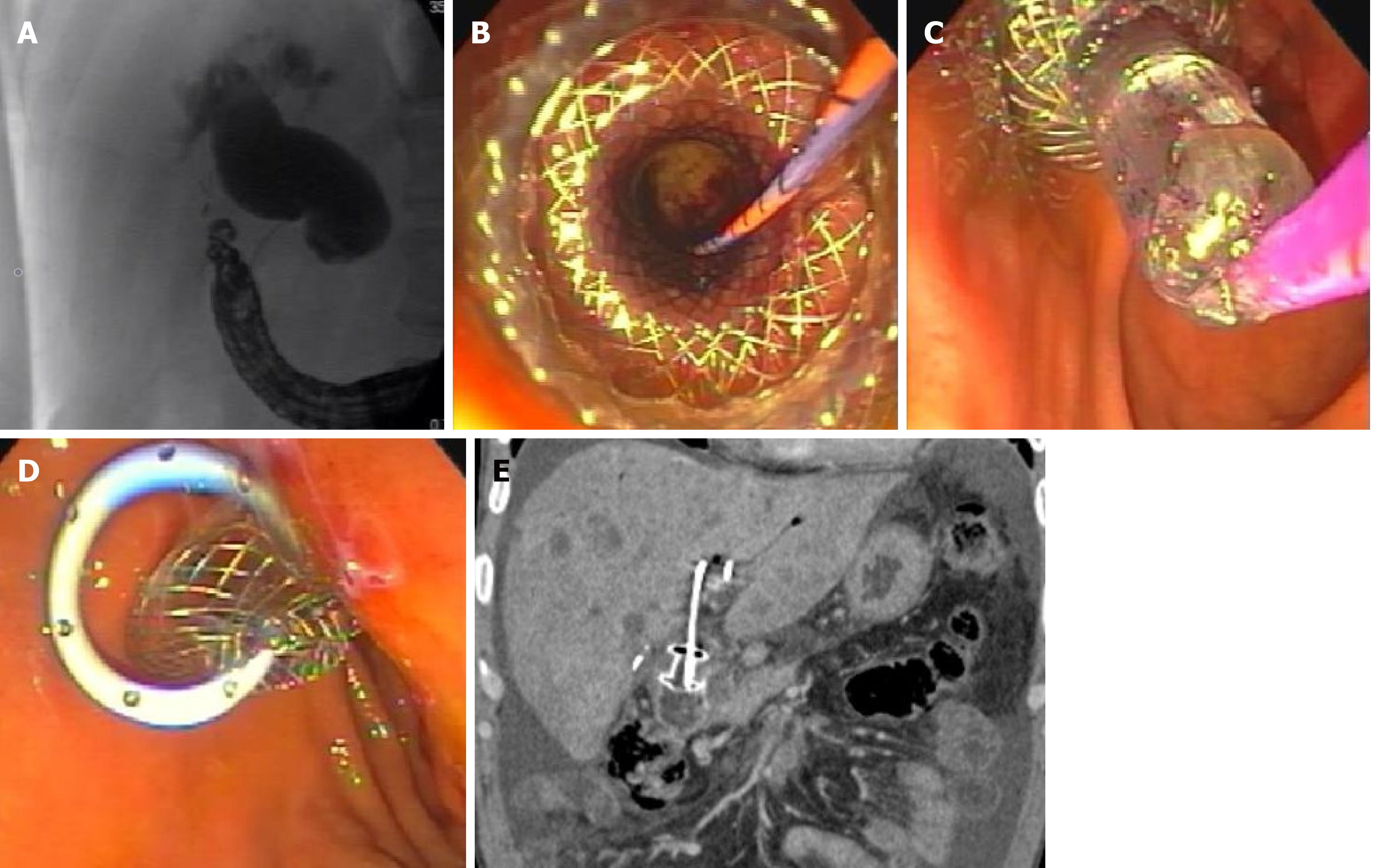
Figure 1 Endoscopic ultrasound-guided choledochoduodenostomy for distal malignant biliary obstruction using an electrocautery-enhanced lumen apposing metal stent.
A: Fluoroscopic image showing a dilated bile duct with distal biliary stricture secondary to pancreas head mass; B: Endoscopic image following lumen-apposing self-expanding metal stent (LAMS) deployment in the common bile duct; C: Balloon dilation of LAMS using a wire-guided balloon; D: Endoscopic image with double pigtail stent through the LAMS in the duodenal bulb; E: Computed tomography coronal image showing choledochoduodenostomy with a double pigtail stent through the LAMS. The proximal end of the double pigtail plastic stent is in the left intrahepatic duct.
- Citation: Pawa R, Pleasant T, Tom C, Pawa S. Endoscopic ultrasound-guided biliary drainage: Are we there yet? World J Gastrointest Endosc 2021; 13(8): 302-318
- URL: https://www.wjgnet.com/1948-5190/full/v13/i8/302.htm
- DOI: https://dx.doi.org/10.4253/wjge.v13.i8.302


