Copyright
©The Author(s) 2021.
World J Gastrointest Endosc. May 16, 2021; 13(5): 155-160
Published online May 16, 2021. doi: 10.4253/wjge.v13.i5.155
Published online May 16, 2021. doi: 10.4253/wjge.v13.i5.155
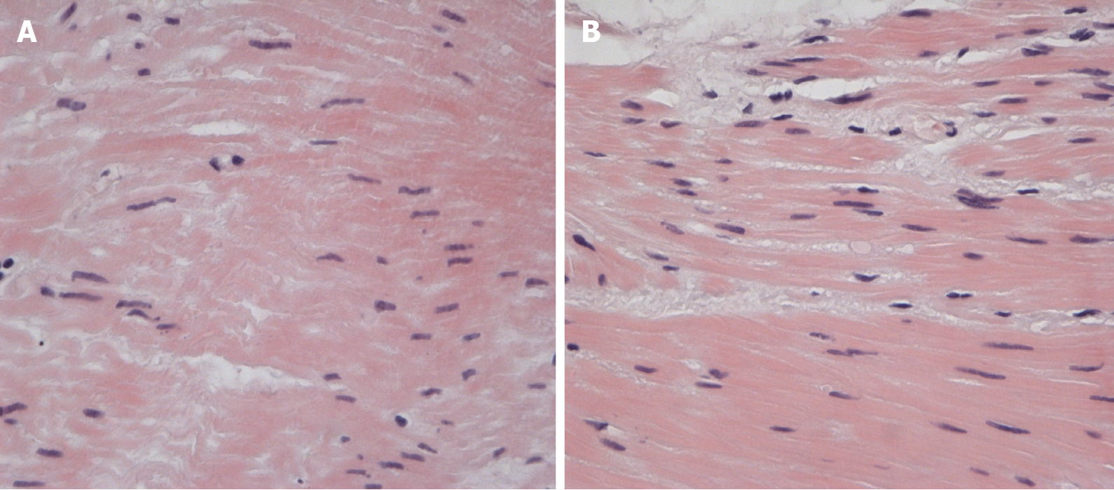
Figure 3 Muscle specimen of the esophagus.
A: Muscle specimen of the upper part of the esophagus. Dystrophic and necrobiotic changes with focal myocytolysis of muscle fibers, hematoxylin-eosin, magnification × 400; B: Muscle specimen of the lower part of the esophagus. Intracellular edema, myocytes of different thicknesses, hematoxylin-eosin, magnification × 400.
- Citation: Smirnov AA, Kiriltseva MM, Lyubchenko ME, Nazarov VD, Botina AV, Burakov AN, Lapin SV. Peroral endoscopic myotomy in a pregnant woman diagnosed with mitochondrial disease: A case report. World J Gastrointest Endosc 2021; 13(5): 155-160
- URL: https://www.wjgnet.com/1948-5190/full/v13/i5/155.htm
- DOI: https://dx.doi.org/10.4253/wjge.v13.i5.155


