Copyright
©The Author(s) 2021.
World J Gastrointest Endosc. Oct 16, 2021; 13(10): 491-501
Published online Oct 16, 2021. doi: 10.4253/wjge.v13.i10.491
Published online Oct 16, 2021. doi: 10.4253/wjge.v13.i10.491
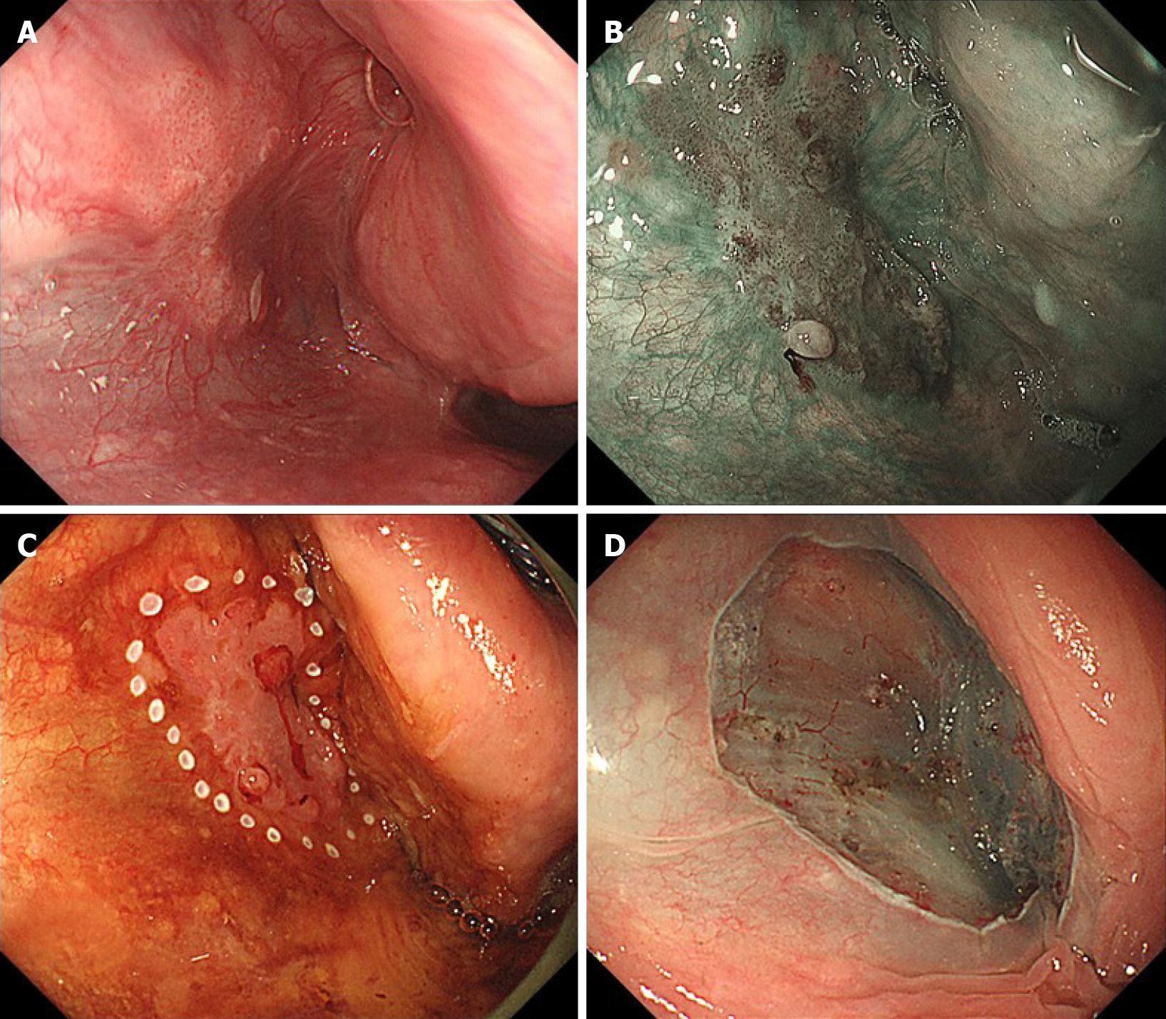
Figure 1 A case of T1 hypopharyngeal cancer located in the left pyriform sinus, detected by gastrointestinal endoscopy.
A: The lesion was recognized as a slightly reddish area under white light image endoscopy; B: The lesion was clearly visualized using narrow-band imaging; C, D: Under general anesthesia, en bloc endoscopic submucosal dissection was successfully completed.
- Citation: Miyamoto H, Naoe H, Morinaga J, Sakisaka K, Tayama S, Matsuno K, Gushima R, Tateyama M, Shono T, Imuta M, Miyamaru S, Murakami D, Orita Y, Tanaka Y. Clinical impact of gastrointestinal endoscopy on the early detection of pharyngeal squamous cell carcinoma: A retrospective cohort study. World J Gastrointest Endosc 2021; 13(10): 491-501
- URL: https://www.wjgnet.com/1948-5190/full/v13/i10/491.htm
- DOI: https://dx.doi.org/10.4253/wjge.v13.i10.491


