Copyright
©The Author(s) 2019.
World J Gastrointest Endosc. May 16, 2019; 11(5): 373-382
Published online May 16, 2019. doi: 10.4253/wjge.v11.i5.373
Published online May 16, 2019. doi: 10.4253/wjge.v11.i5.373
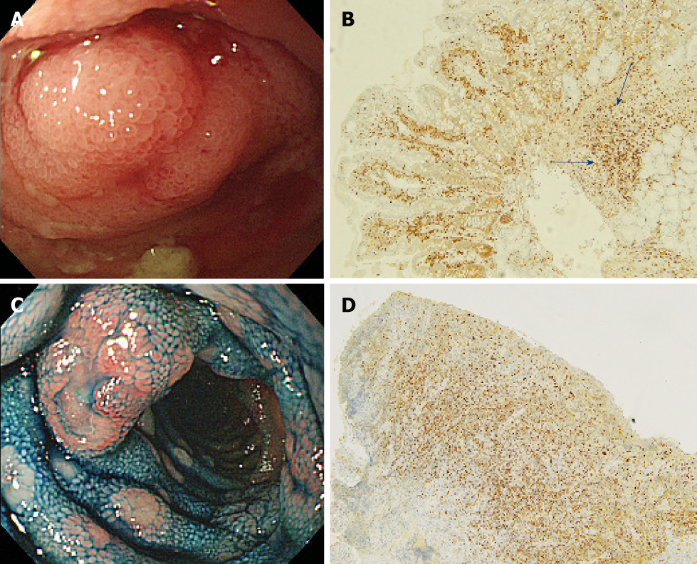
Figure 4 Endoscopic and pathological images of malignant lymphoma without white villi appearance.
A, B: Representative endoscopic and pathological images of malignant lymphoma without white villi appearance (Follicular lymphoma) [A: White light image shows enlarged villi with stenosis, without white villi appearance in the ileum (with stenosis); B: Pathological image is showing the lymphoma cells sparsely infiltrating the villi, some lymphoma cells present in the deep mucosa (blue arrow), immunohistochemical staining for bcl-2, ×10]; C, D: Representative endoscopic and pathological images of malignant lymphoma without white villi appearance (Mantle cell lymphoma) (C: White light image with indigo carmine staining shows multiple polyposis with ulceration, without white villi appearance in the ileum; D: Pathological image is showing the lymphoma cells infiltrating the mucosa without an intact epithelium, immunohistochemical staining for cyclin D1, ×10).
- Citation: Horie T, Hosoe N, Takabayashi K, Hayashi Y, Kamiya KJL, Miyanaga R, Mizuno S, Fukuhara K, Fukuhara S, Naganuma M, Shimoda M, Ogata H, Kanai T. Endoscopic characteristics of small intestinal malignant tumors observed by balloon-assisted enteroscopy. World J Gastrointest Endosc 2019; 11(5): 373-382
- URL: https://www.wjgnet.com/1948-5190/full/v11/i5/373.htm
- DOI: https://dx.doi.org/10.4253/wjge.v11.i5.373


