Copyright
©The Author(s) 2019.
World J Gastrointest Endosc. Mar 16, 2019; 11(3): 174-192
Published online Mar 16, 2019. doi: 10.4253/wjge.v11.i3.174
Published online Mar 16, 2019. doi: 10.4253/wjge.v11.i3.174
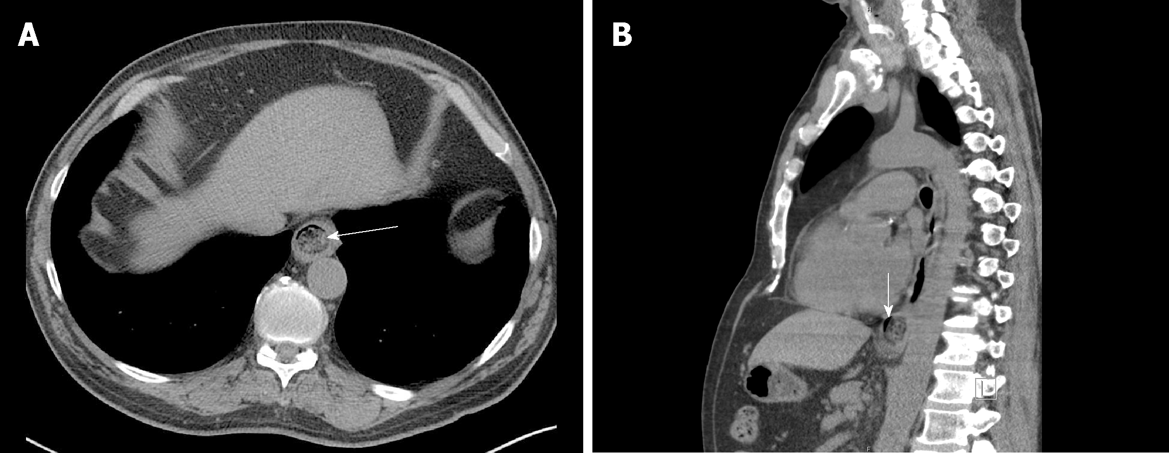
Figure 2 Computed tomography revealing an esophageal food impaction.
A: Axial tomogram revealing a food bolus (arrow) in the distal esophagus; B: Sagittal tomogram reveals a sliver of space around the bolus (vertical arrow) suggestive of an opportunity to wedge in the endoscope and employ the push technique or to pass a guidewire (e.g., for the balloon dilation technique).
- Citation: Fung BM, Sweetser S, Wong Kee Song LM, Tabibian JH. Foreign object ingestion and esophageal food impaction: An update and review on endoscopic management. World J Gastrointest Endosc 2019; 11(3): 174-192
- URL: https://www.wjgnet.com/1948-5190/full/v11/i3/174.htm
- DOI: https://dx.doi.org/10.4253/wjge.v11.i3.174


