Copyright
©The Author(s) 2018.
World J Gastrointest Endosc. May 16, 2018; 10(5): 99-108
Published online May 16, 2018. doi: 10.4253/wjge.v10.i5.99
Published online May 16, 2018. doi: 10.4253/wjge.v10.i5.99
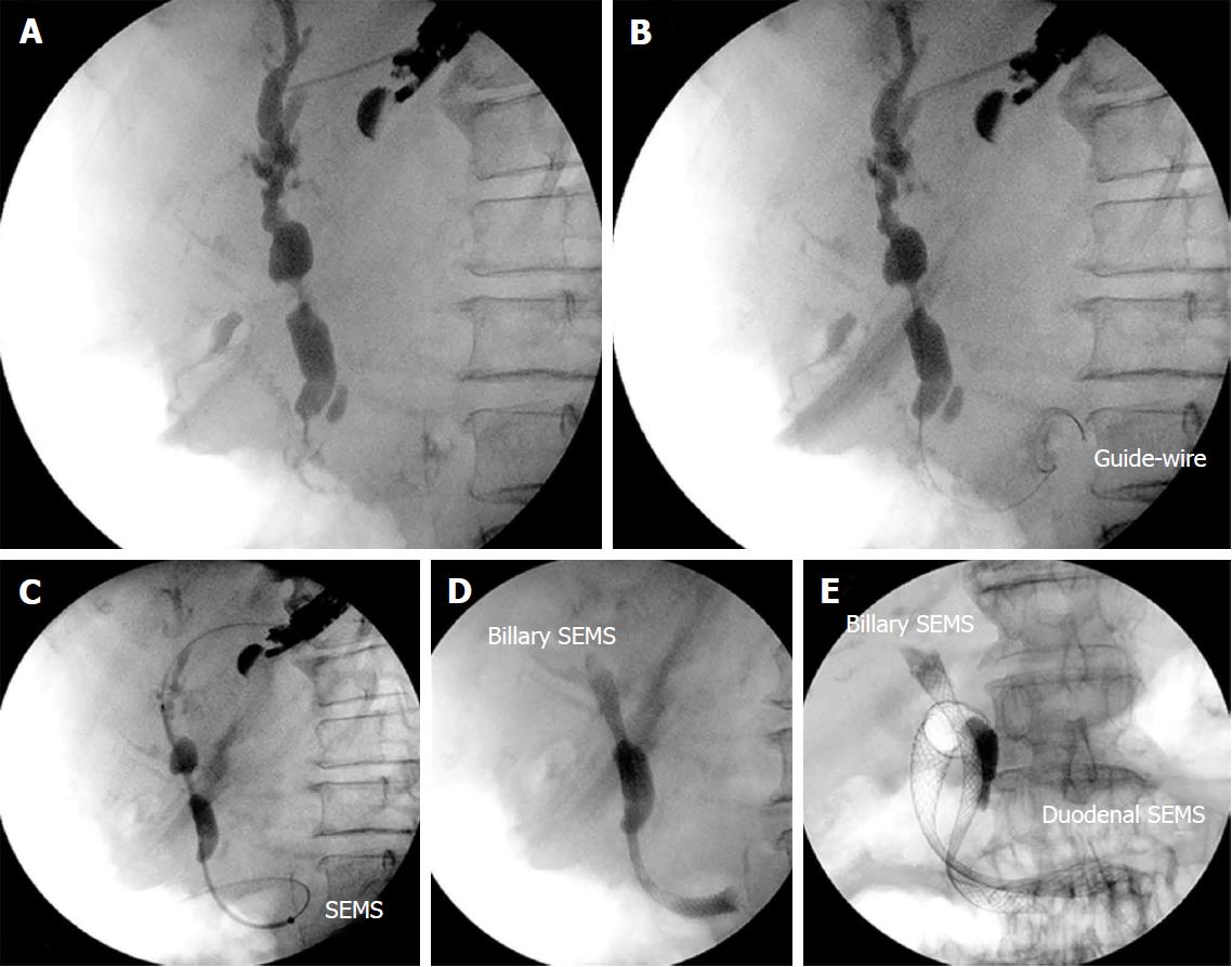
Figure 3 Patient with duodenal stenosis due to a pancreatic carcinoma.
A: Endosonography (EUS)-guided cholangiography; B: Insertion of the guidewire through the duodenal major papilla and positioning in the duodenum; C: Anterograde insertion of the self-expandable metallic stents (SEMS) through the gastric wall across the duodenal major papilla and its positioning in the duodenum; D: Deployment of the SEMS; E: Insertion of the duodenal SEMS. SEMS: Self-expandable metallic stents.
- Citation: Ardengh JC, Lopes CV, Kemp R, dos Santos JS. Different options of endosonography-guided biliary drainage after endoscopic retrograde cholangio-pancreatography failure. World J Gastrointest Endosc 2018; 10(5): 99-108
- URL: https://www.wjgnet.com/1948-5190/full/v10/i5/99.htm
- DOI: https://dx.doi.org/10.4253/wjge.v10.i5.99


