Copyright
©The Author(s) 2018.
World J Gastrointest Endosc. Feb 16, 2018; 10(2): 56-68
Published online Feb 16, 2018. doi: 10.4253/wjge.v10.i2.56
Published online Feb 16, 2018. doi: 10.4253/wjge.v10.i2.56
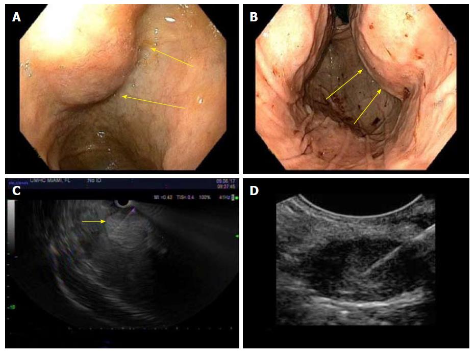
Figure 2 Endoscopic ultrasonography (EUS) diagnosis of primary liver tumors.
A and B: Indentations seen in the duodenal bulb and stomach, from large lesions in the right and left lobes of the liver; C: On EUS, the entire liver tissue was seen replaced by numerous hyperechoic lesions of medium size, causing hepatomegaly and hence indentations. Fine needle aspiration (FNA) of a prominent and accessible hyperechoic lesion obtained, which was diagnostic of neuroendocrine tumor; D: In another patient, EUS-FNA of solitary hypoechoic liver lesion obtained, which diagnosed hepatocellular carcinoma.
- Citation: Girotra M, Soota K, Dhaliwal AS, Abraham RR, Garcia-Saenz-de-Sicilia M, Tharian B. Utility of endoscopic ultrasound and endoscopy in diagnosis and management of hepatocellular carcinoma and its complications: What does endoscopic ultrasonography offer above and beyond conventional cross-sectional imaging? World J Gastrointest Endosc 2018; 10(2): 56-68
- URL: https://www.wjgnet.com/1948-5190/full/v10/i2/56.htm
- DOI: https://dx.doi.org/10.4253/wjge.v10.i2.56


