Copyright
©The Author(s) 2018.
World J Gastrointest Endosc. Nov 16, 2018; 10(11): 367-377
Published online Nov 16, 2018. doi: 10.4253/wjge.v10.i11.367
Published online Nov 16, 2018. doi: 10.4253/wjge.v10.i11.367
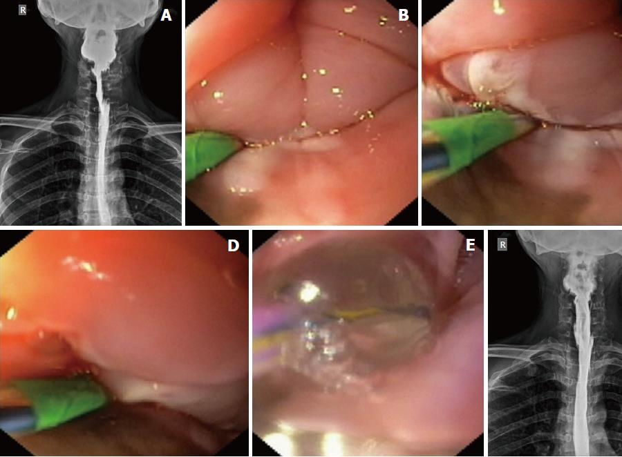
Figure 3 Near-total stricture in case 1 and its endoscopic management.
A: The esophagogram after dilatation in case 1 showed a residual eccentric stricture, suggestive of persistent adhesions in the post-cricoid space and left piriform sinus; B-D: Serial endoscopic images of electroincision procedure in case 1, as the wire-guided sphincterotome was progressively moving towards the left pharyngeal wall after cutting the adhesions in the post-cricoid space and left piriform sinus; E: After electro-incision, a dilatation was given with the dilating balloon placed in the left piriform sinus; F: Follow-up esophagogram showing the completely opened-up stricture along its entire length.
- Citation: Dhaliwal HS, Kumar N, Siddappa PK, Singh R, Sekhon JS, Masih J, Abraham J, Garg S. Tight near-total corrosive strictures of the proximal esophagus with concomitant involvement of the hypopharynx: Flexible endoscopic management using a novel technique. World J Gastrointest Endosc 2018; 10(11): 367-377
- URL: https://www.wjgnet.com/1948-5190/full/v10/i11/367.htm
- DOI: https://dx.doi.org/10.4253/wjge.v10.i11.367


