















©The Author(s) 2018.
World J Gastrointest Endosc. Jan 16, 2018; 10(1): 10-15
Published online Jan 16, 2018. doi: 10.4253/wjge.v10.i1.10
Published online Jan 16, 2018. doi: 10.4253/wjge.v10.i1.10
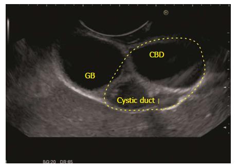
Figure 14 The gall bladder imaging is done from descending duodenum.
The hepatoduodenal ligament is identified between the probe and liver (shown in dotted yellow area). The CBD, the cystic duct and the gall bladder are visualized on the under surface of liver. CBD: Common bile duct; GB: Gall bladder.
- Citation: Sharma M, Somani P, Sunkara T. Imaging of gall bladder by endoscopic ultrasound. World J Gastrointest Endosc 2018; 10(1): 10-15
- URL: https://www.wjgnet.com/1948-5190/full/v10/i1/10.htm
- DOI: https://dx.doi.org/10.4253/wjge.v10.i1.10