Copyright
©The Author(s) 2018.
World J Gastrointest Endosc. Jan 16, 2018; 10(1): 10-15
Published online Jan 16, 2018. doi: 10.4253/wjge.v10.i1.10
Published online Jan 16, 2018. doi: 10.4253/wjge.v10.i1.10
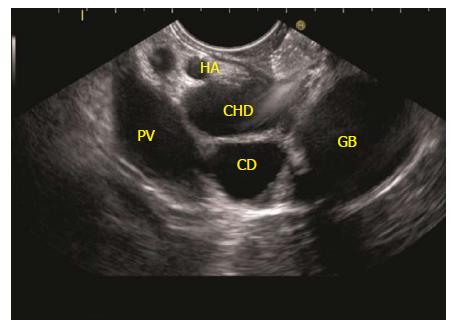
Figure 12 The imaging is done from duodenal bulb and the portal vein is identified going from 5 o’clock position to 10 o’clock position in a long axis.
The CHD is identified between the probe and portal vein. The CHD is followed up by anticlockwise rotation and the continuity into cystic duct and gall bladder is seen. CHD: Common hepatic duct; PV: Portal vein; GB: Gall bladder.
- Citation: Sharma M, Somani P, Sunkara T. Imaging of gall bladder by endoscopic ultrasound. World J Gastrointest Endosc 2018; 10(1): 10-15
- URL: https://www.wjgnet.com/1948-5190/full/v10/i1/10.htm
- DOI: https://dx.doi.org/10.4253/wjge.v10.i1.10


