Copyright
©The Author(s) 2023.
World J Gastrointest Endosc. Mar 16, 2023; 15(3): 114-121
Published online Mar 16, 2023. doi: 10.4253/wjge.v15.i3.114
Published online Mar 16, 2023. doi: 10.4253/wjge.v15.i3.114
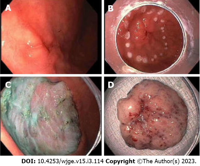
Figure 1 Endoscopic submucosal dissection of a type 0-IIc lesion found in the antrum.
A: Lesion noted in the antrum; B: Lesion marked for endoscopic submucosal dissection (ESD); C: Lesion removed successfully with ESD; D: Removed specimen, pathology returned as well-differentiated adenocarcinoma with no evidence of malignancy at the margins and no lymph node invasion (courtesy of Dr. Makoto Nishimura).
- Citation: Park E, Nishimura M, Simoes P. Endoscopic advances in the management of gastric cancer and premalignant gastric conditions. World J Gastrointest Endosc 2023; 15(3): 114-121
- URL: https://www.wjgnet.com/1948-5190/full/v15/i3/114.htm
- DOI: https://dx.doi.org/10.4253/wjge.v15.i3.114


