Copyright
©The Author(s) 2023.
World J Gastrointest Endosc. Oct 16, 2023; 15(10): 614-622
Published online Oct 16, 2023. doi: 10.4253/wjge.v15.i10.614
Published online Oct 16, 2023. doi: 10.4253/wjge.v15.i10.614
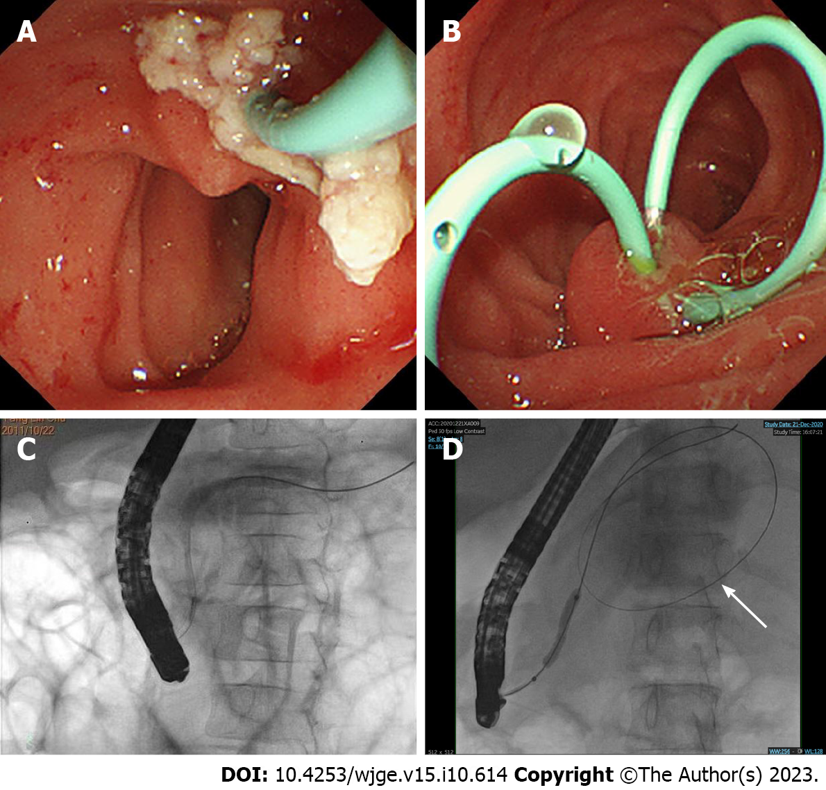
Figure 1 Endoscopic retrograde cholangiopancreatography procedures.
A: Endoscopic view of stone extraction; B: Endoscopic view showing that two pancreatic stents were placed after stone extraction; C: Fluoroscopic view of endoscopic retrograde cholangiopancreatography showing the dilated and tortuous pancreatic duct; D: Fluoroscopic view of pancreatic duct stricture dilation performed by balloon manipulation. White arrow shows the guide wire coiled inside the pseudocyst.
- Citation: Yang KH, Zeng JQ, Ding S, Zhang TA, Wang WY, Zhang JY, Wang L, Xiao J, Gong B, Deng ZH. Efficacy and safety of endoscopic retrograde cholangiopancreatography in recurrent pancreatitis of pediatric asparaginase-associated pancreatitis. World J Gastrointest Endosc 2023; 15(10): 614-622
- URL: https://www.wjgnet.com/1948-5190/full/v15/i10/614.htm
- DOI: https://dx.doi.org/10.4253/wjge.v15.i10.614


