Copyright
©The Author(s) 2020.
World J Gastrointest Endosc. Mar 16, 2020; 12(3): 111-118
Published online Mar 16, 2020. doi: 10.4253/wjge.v12.i3.111
Published online Mar 16, 2020. doi: 10.4253/wjge.v12.i3.111
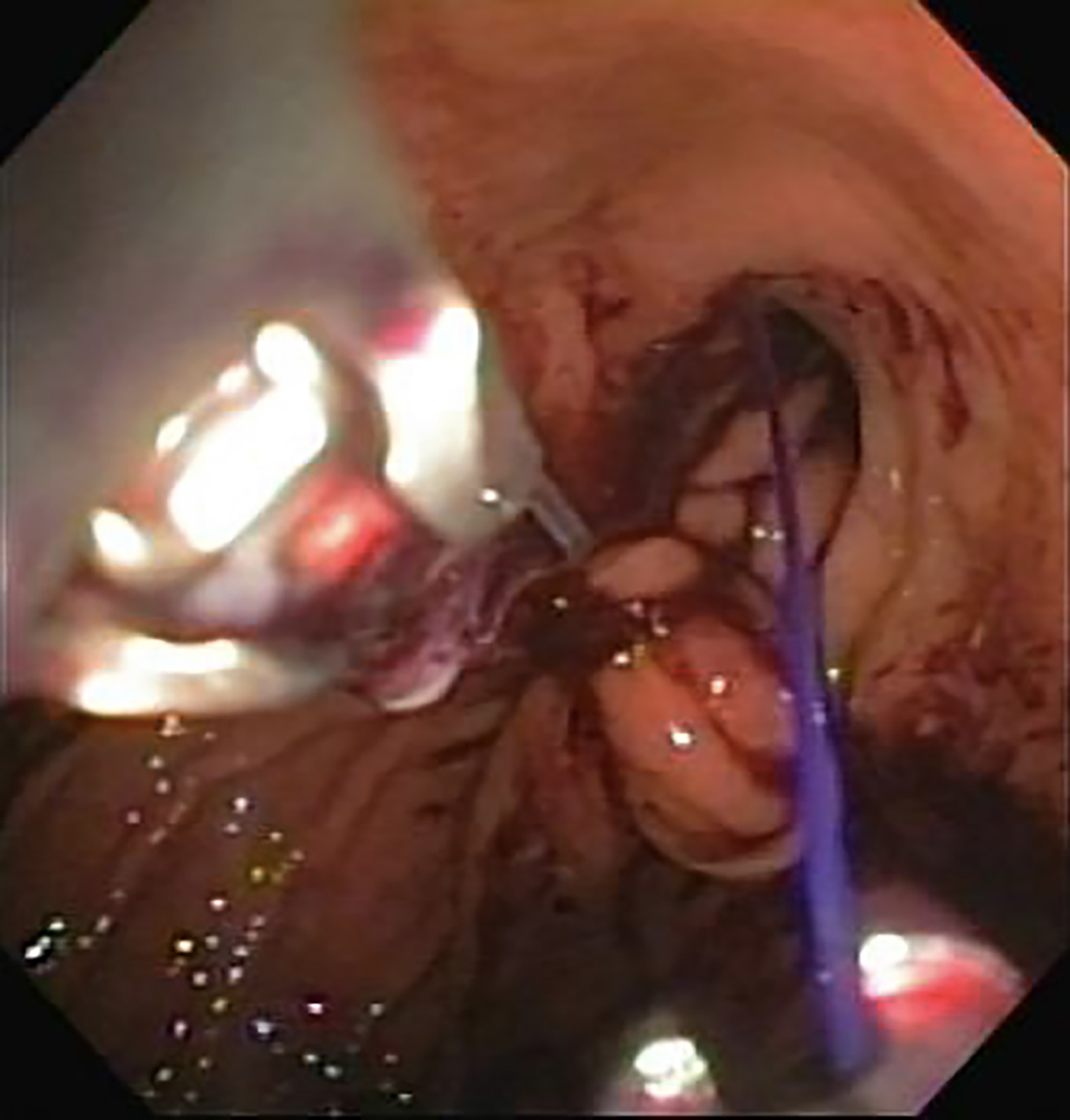
Figure 1 Endoscopic suture placement during endoscopic sleeve.
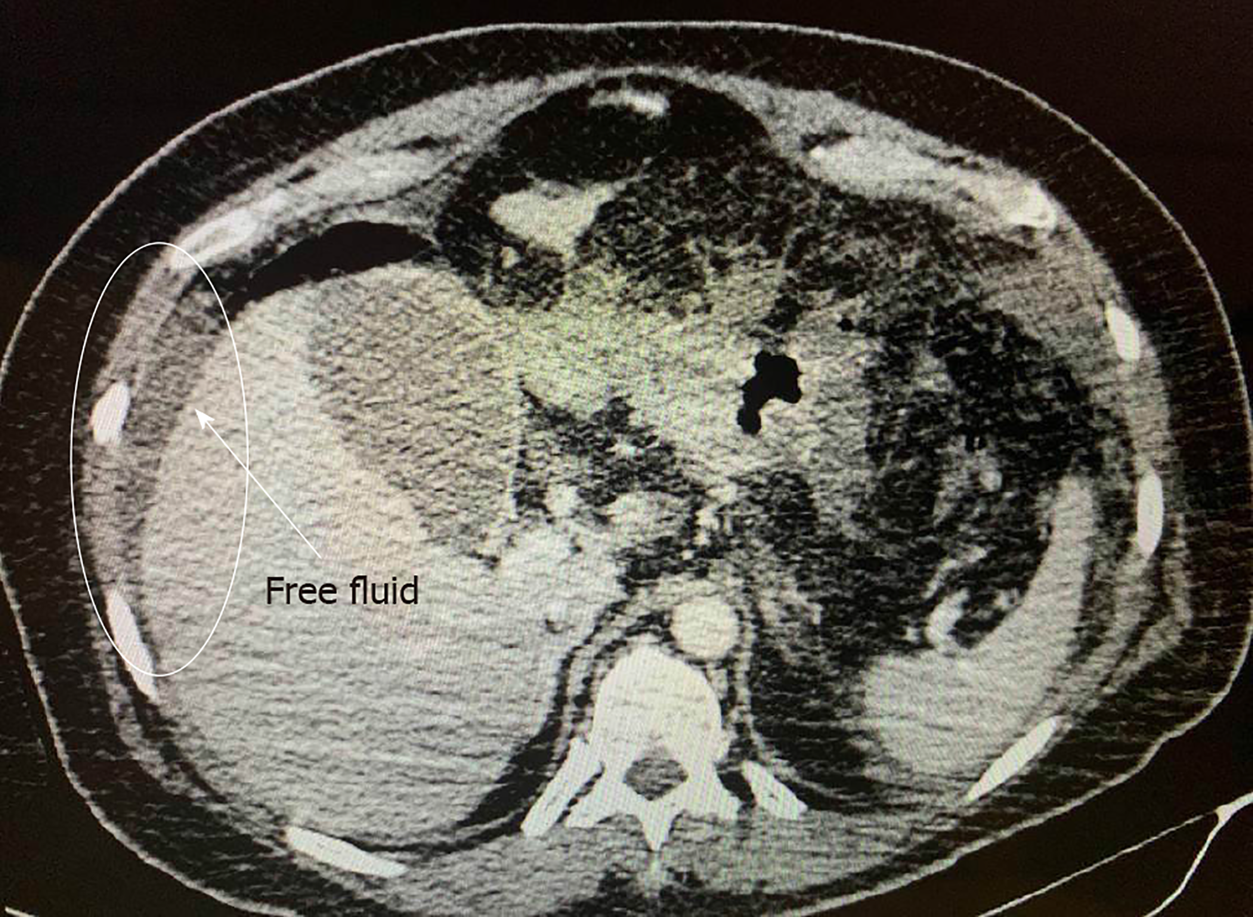
Figure 2 Computerized tomography showing free fluid in the abdominal cavity.
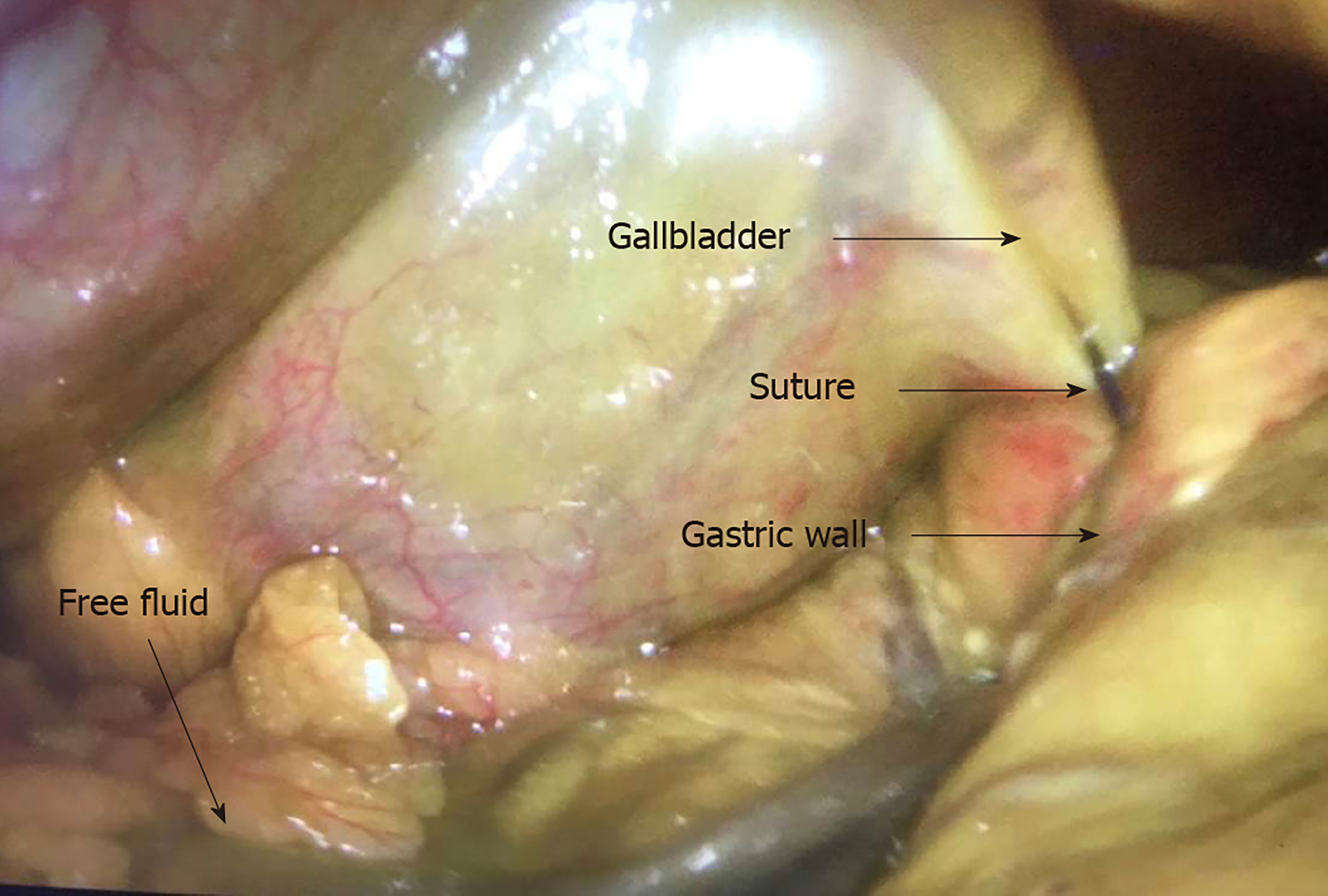
Figure 3 Laparoscopic visualization showing bile ascites, the gallbladder, the stomach, and a suture between the gallbladder and the stomach.
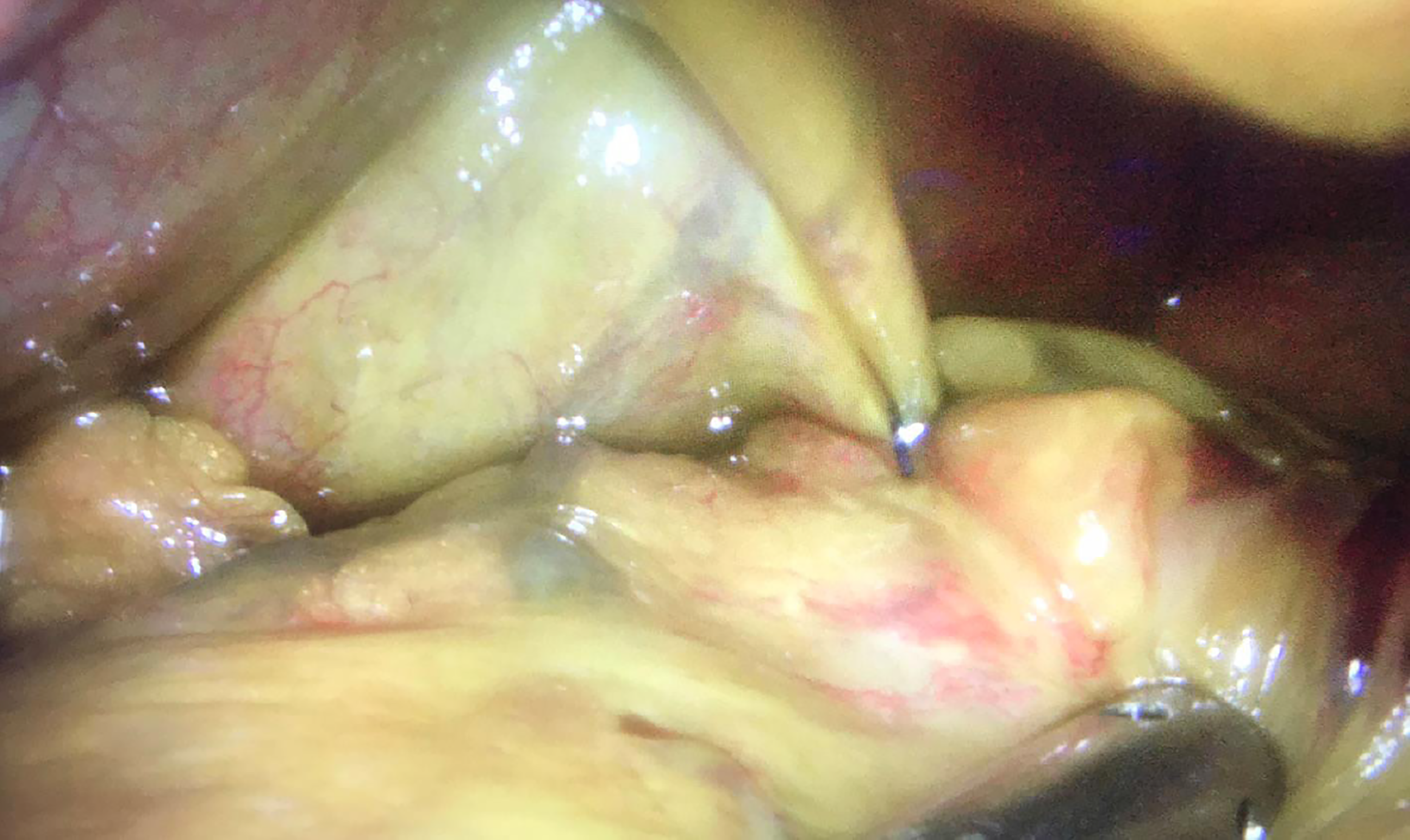
Figure 4 Laparoscopic visualization of a suture in the gallbladder after aspiration of bile ascites.
- Citation: de Siqueira Neto J, de Moura DTH, Ribeiro IB, Barrichello SA, Harthorn KE, Thompson CC. Gallbladder perforation due to endoscopic sleeve gastroplasty: A case report and review of literature. World J Gastrointest Endosc 2020; 12(3): 111-118
- URL: https://www.wjgnet.com/1948-5190/full/v12/i3/111.htm
- DOI: https://dx.doi.org/10.4253/wjge.v12.i3.111


