Copyright
©The Author(s) 2020.
World J Gastrointest Endosc. Dec 16, 2020; 12(12): 521-531
Published online Dec 16, 2020. doi: 10.4253/wjge.v12.i12.521
Published online Dec 16, 2020. doi: 10.4253/wjge.v12.i12.521
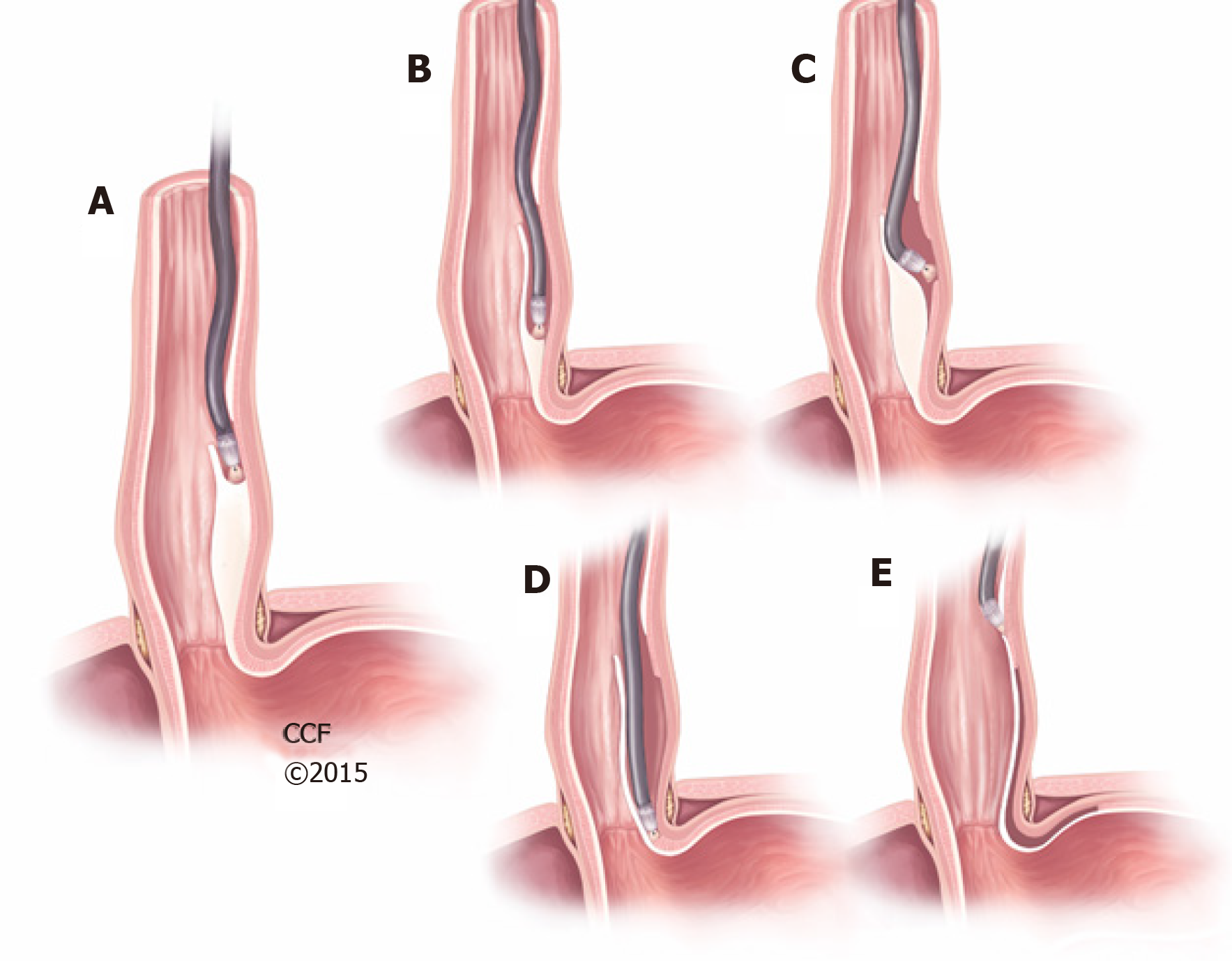
Figure 1 Peroral endoscopic myotomy.
A: Submucosal injection in the mid esophagus to create a submucosal bleb, followed by a mucosal incision to enter the submucosal space; B: Create a submucosal tunnel along the esophageal length; C and D: Esophagogastric myotomy; E: Closure of submucosal entry point with endoscopic clips. Reprinted with permission, Cleveland Clinic Center for Medical Art & Photography© 2015-2020. All Rights Reserved.
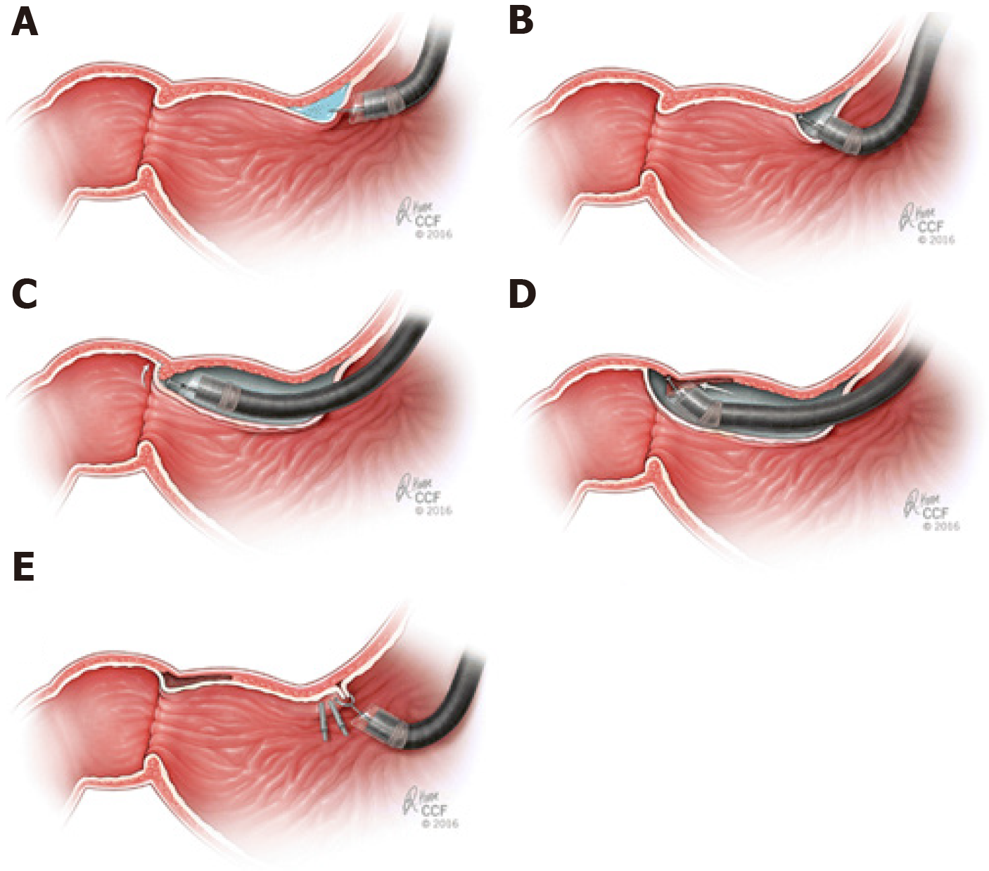
Figure 2 Per-oral Pyloromyotomy.
A: Submucosal injection with a coloring agent proximal to the pylorus to create a submucosal bleb; B: Mucosotomy; C: Dissection of the submucosa until the pylorus is identified; D: Pyloromyotomy performed in a distal to proximal fashion; E: Closure of the mucosotomy by endoscopic clips. Reprinted with permission, Cleveland Clinic Center for Medical Art & Photography© 2015-2020. All Rights Reserved.
Figure 3 Submucosal tunneling endoscopic septum division.
A and B: Mucosal incision proximal to the septum followed by creation of a submucosal tunnel; C: Cricopharyngeal muscle fibers dissected to the bottom of the diverticulum; D and E: Closure of the entry site with endoscopic clips. Reprinted with permission, Cleveland Clinic Center for Medical Art & Photography© 2015-2020. All Rights Reserved.
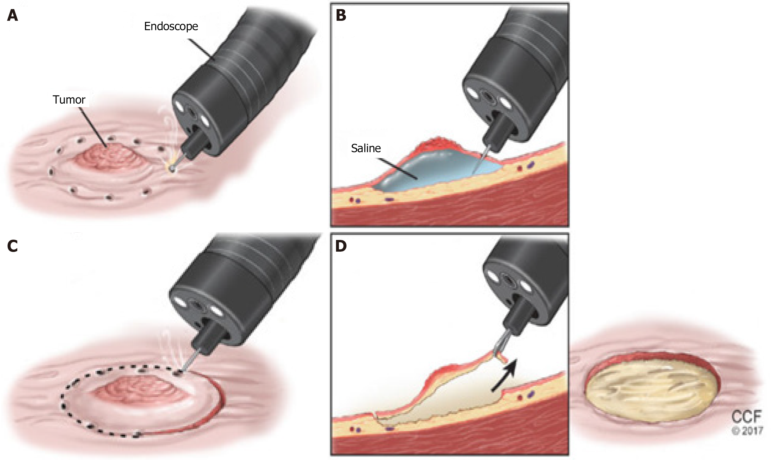
Figure 4 Endoscopic submucosal dissection.
A: Lesion marked with cautery; B: Submucosal injection to create a bleb; C: Circumferential mucosal incision; D: Followed by submucosal dissection until the lesion is removed. Reprinted with permission, Cleveland Clinic Center for Medical Art & Photography© 2015-2020. All Rights Reserved.
- Citation: Shanbhag AB, Thota PN, Sanaka MR. Recent advances in third space or intramural endoscopy. World J Gastrointest Endosc 2020; 12(12): 521-531
- URL: https://www.wjgnet.com/1948-5190/full/v12/i12/521.htm
- DOI: https://dx.doi.org/10.4253/wjge.v12.i12.521


