Copyright
©The Author(s) 2020.
World J Clin Cases. Nov 6, 2020; 8(21): 5149-5158
Published online Nov 6, 2020. doi: 10.12998/wjcc.v8.i21.5149
Published online Nov 6, 2020. doi: 10.12998/wjcc.v8.i21.5149
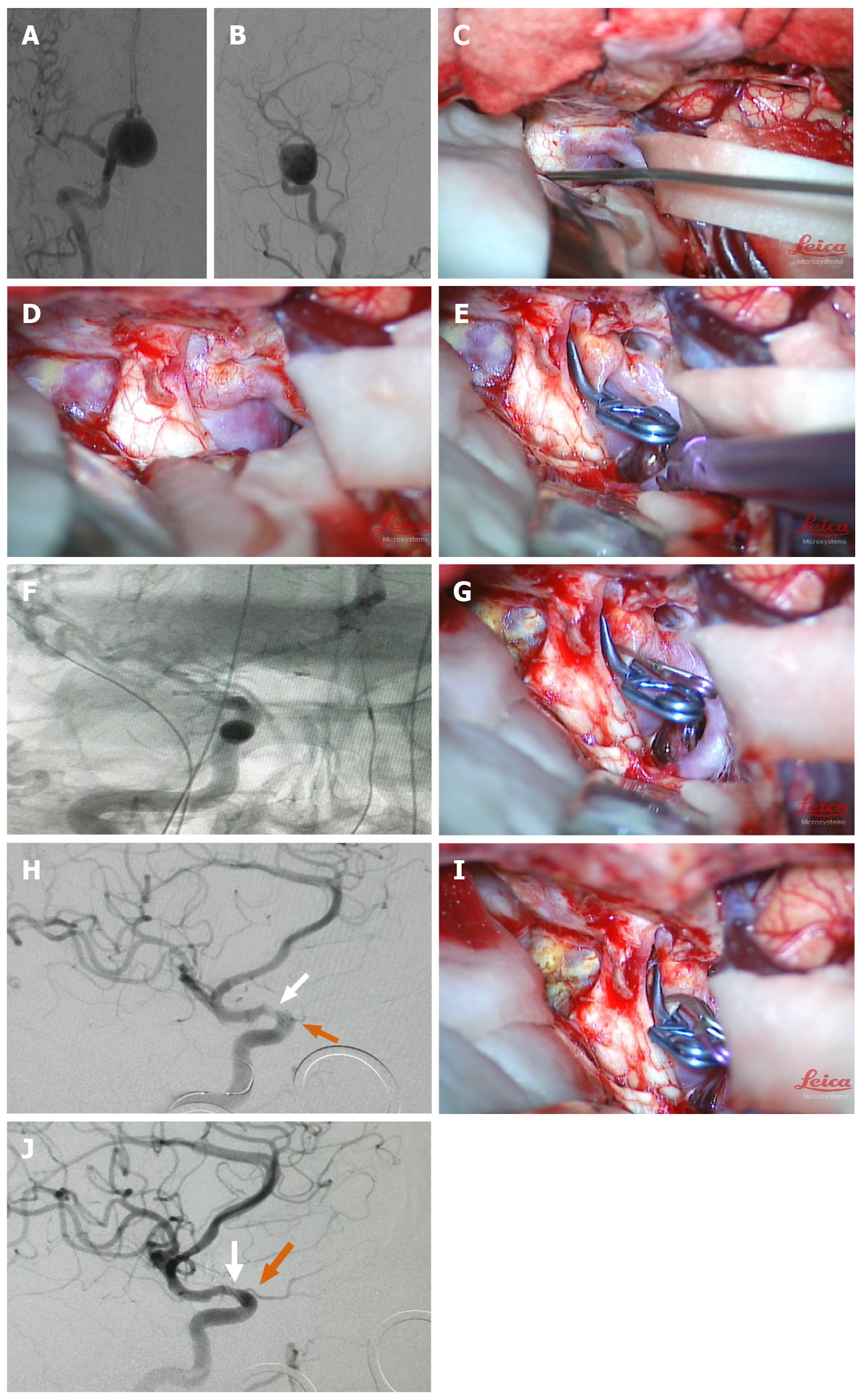
Figure 1 Preoperative, intraoperative and postoperative imaging of a 56-year-old female patient with aneurysm of the right ophthalmic segment.
A and B: Pre-operative digital subtraction angiography (DSA) in anteroposterior and lateral views: The stained aneurysm was visible in the dextral internal carotid artery (ICA) ophthalmic artery segment, maximal diameter (25.3) mm, neck size (7.5 mm), and dome-neck ratio of 3.4; C and D: The optic nerve was compressed by the giant aneurysm; removal of the anterior clinoid process and dissociation of the optic nerve from the aneurysm neck to sufficiently reveal the aneurysm neck; E and F: Placement of the first clip; first intraoperative DSA showed that the aneurysm had been clipped, but a small portion of residual aneurysm neck was visualized; G and H: Supplementary miniature clip was placed during the operation; second intraoperative DSA displayed that the aneurysm had been completely clipped; however, stenosis occurred in the ICA (white arrow) with poor visualization of the optic nerve (orange arrow) and poor visualization of the distal branch vessel of the middle cerebral artery (MCA); I and J: A miniature clip with angular tips was used to replace the original clip. Third intraoperative DSA showed that the aneurysm was completely clipped. ICA stenosis and ophthalmic artery stenosis were relieved (white arrow), and the distal branch vessel of the MCA was improved (orange arrow).
- Citation: Zhang N, Xin WQ. Application of hybrid operating rooms for clipping large or giant intracranial carotid-ophthalmic aneurysms. World J Clin Cases 2020; 8(21): 5149-5158
- URL: https://www.wjgnet.com/2307-8960/full/v8/i21/5149.htm
- DOI: https://dx.doi.org/10.12998/wjcc.v8.i21.5149


