Copyright
©The Author(s) 2019.
World J Clin Cases. Dec 6, 2019; 7(23): 4004-4010
Published online Dec 6, 2019. doi: 10.12998/wjcc.v7.i23.4004
Published online Dec 6, 2019. doi: 10.12998/wjcc.v7.i23.4004
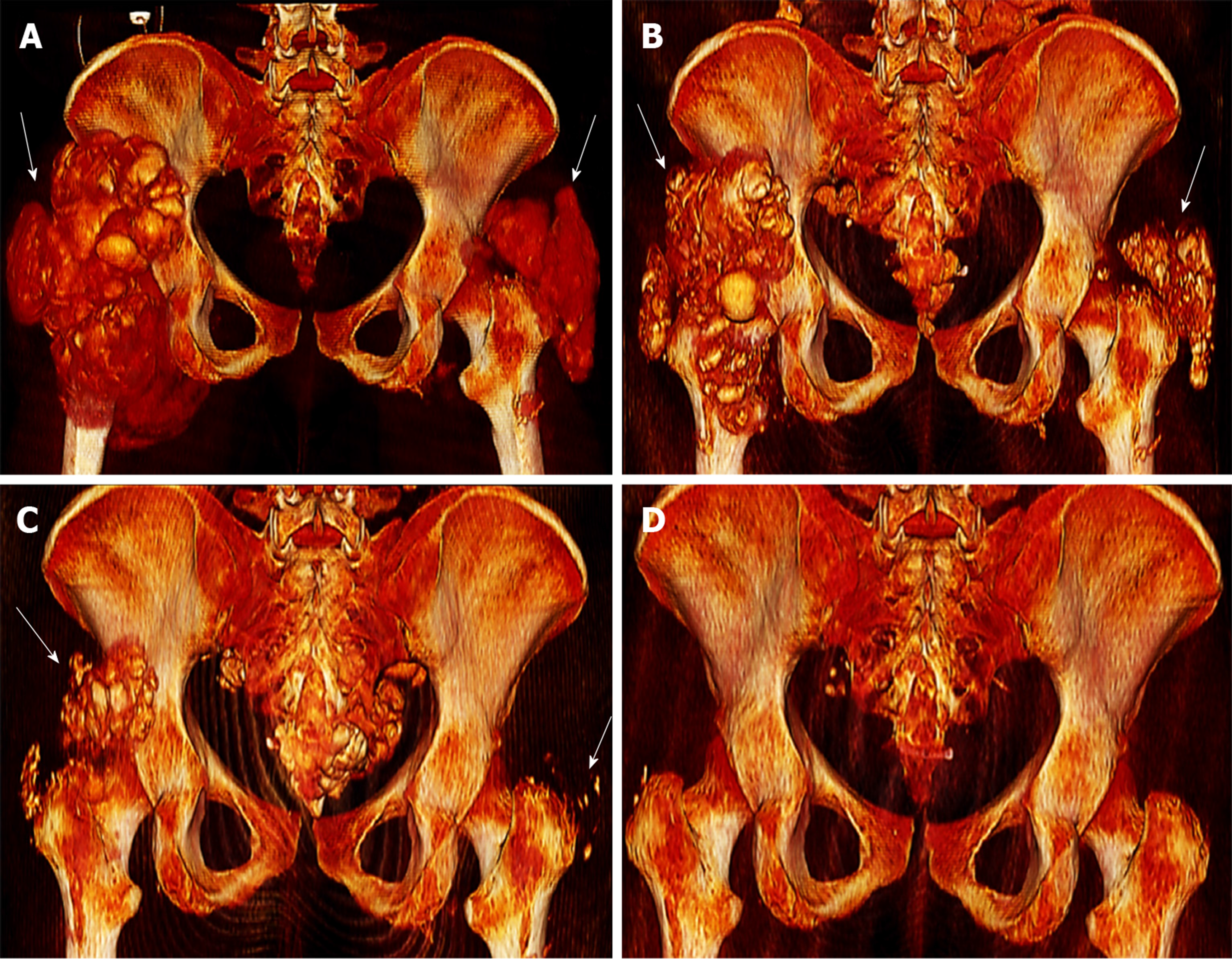
Figure 2 3D reconstruction of computed tomography scan images.
A: Initial diagnosis; B: At 2 mo; C: At 4 mo; D: At 11 mo. Computed tomography scan reconstructions show 3D spatial expansion of tumoral calcinosis lesions of the trochanter major region (arrows) depicting their severity at initial diagnosis (A) and their continuous remission thereafter at 2 mo (B), 4 mo (C) and finally 11 mo (D) after the initial diagnosis at which point complete remission was achieved (all images represent dorsal pelvic view).
- Citation: Westermann L, Isbell LK, Breitenfeldt MK, Arnold F, Röthele E, Schneider J, Widmeier E. Recuperation of severe tumoral calcinosis in a dialysis patient: A case report. World J Clin Cases 2019; 7(23): 4004-4010
- URL: https://www.wjgnet.com/2307-8960/full/v7/i23/4004.htm
- DOI: https://dx.doi.org/10.12998/wjcc.v7.i23.4004


