Published online Oct 6, 2019. doi: 10.12998/wjcc.v7.i19.3132
Peer-review started: March 26, 2019
First decision: May 31, 2019
Revised: June 25, 2019
Accepted: July 3, 2019
Article in press: July 3, 2019
Published online: October 6, 2019
Parathyroid adenoma (PTA) is known as an adenomatous hyperparathyroidism syndrome. At earlier times, the major symptoms of this disease included high blood calcium and low phosphorus. PTA is a benign neuroendocrine neoplasm. We have reviewed the literature and found that it is rare for patients with hyperparathyroidism to have benign tumors with multiple organs at the same time. This report describes a patient with a PTA and four nonfunctional adenomas.
We report a case of primary hyperparathyroidism in a 39-year-old woman with multiple organ tumors. The patient was admitted to hospital because of hypercalcemia. Laboratory, imaging, and histological examinations confirmed a left parathyroid neoplasm. Right thyroid adenoma was discovered during hospitalization. She had a medical history of uterine fibroids, right benign mammary gland tumor, and meningioma. The patient recovered after surgical and conservative treatments.
Primary hyperparathyroidism with multiple organ tumors is uncommon, and further studies should be conducted to determine if there is genetic heterogeneity.
Core tip: Simultaneous occurrence of multiple tumors is rare. This article reports a patient who was admitted to hospital with hypercalcemia and finally diagnosed with a parathyroid adenoma via surgical pathology. The patient also had uterine fibroids, benign mammary gland tumor, and meningioma. After diagnosis, these tumors were all removed.
- Citation: Hui CC, Zhang X, Sun JR, Deng DT. Primary hyperparathyroidism in a woman with multiple tumors: A case report. World J Clin Cases 2019; 7(19): 3132-3137
- URL: https://www.wjgnet.com/2307-8960/full/v7/i19/3132.htm
- DOI: https://dx.doi.org/10.12998/wjcc.v7.i19.3132
A single person with tumors occurring in multiple organs is rare. Endocrine tumor refers to a series of tumors that not only has the characteristics of a tumor but also has endocrine function. Based on its endocrine function, it can be divided into functional and non-functional.
Here, we report the case of a patient with tumors in multiple organs. Among these, only the parathyroid tumor had endocrine function. Therefore, we postulated that the patient was genetically heterogeneous, which would lead to the emergence of such multiple types of tumors.
A 39-year-old woman attended a local hospital due to abdominal distension, abdominal pain with asthenia, and anorexia.
After abdominal subtotal hysterectomy for multiple myomata of uterus, the patient experienced recurrent abdominal pain and distension. During the next 10 d, these symptoms gradually worsened.
She underwent surgery for a meningeal tumor 4 years ago and resection of a right breast benign tumor 6 mo ago. After subtotal hysterectomy, the patient developed an epileptic seizure in the anesthetic recovery room.
Physical examination on admission revealed blood pressure of 15.8/10.7 mmHg, resting pulse of 90 times/min, body mass index of 22.4 kg/m2, and tenderness beneath the xiphoid process.
Laboratory tests showed increased calcium 4.21 mmol/L, depressed phosphorus 0.85 mmol/L, and elevated urine calcium 8.32 mmol/24 h (reference range 2.50-7.50). The plasma level of parathyroid hormone (PTH) was elevated to 492.00 pg/mL (reference range 10.00-69.00). A diagnosis of hyperparathyroidism was established. Her sex hormone levels were normal, serum 25-hydroxyvitamin D concentration was decreased to 7.0 ng/mL (normal range 30-100), and serum calcitonin was 5.62 pg/mL (normal level < 11.5).
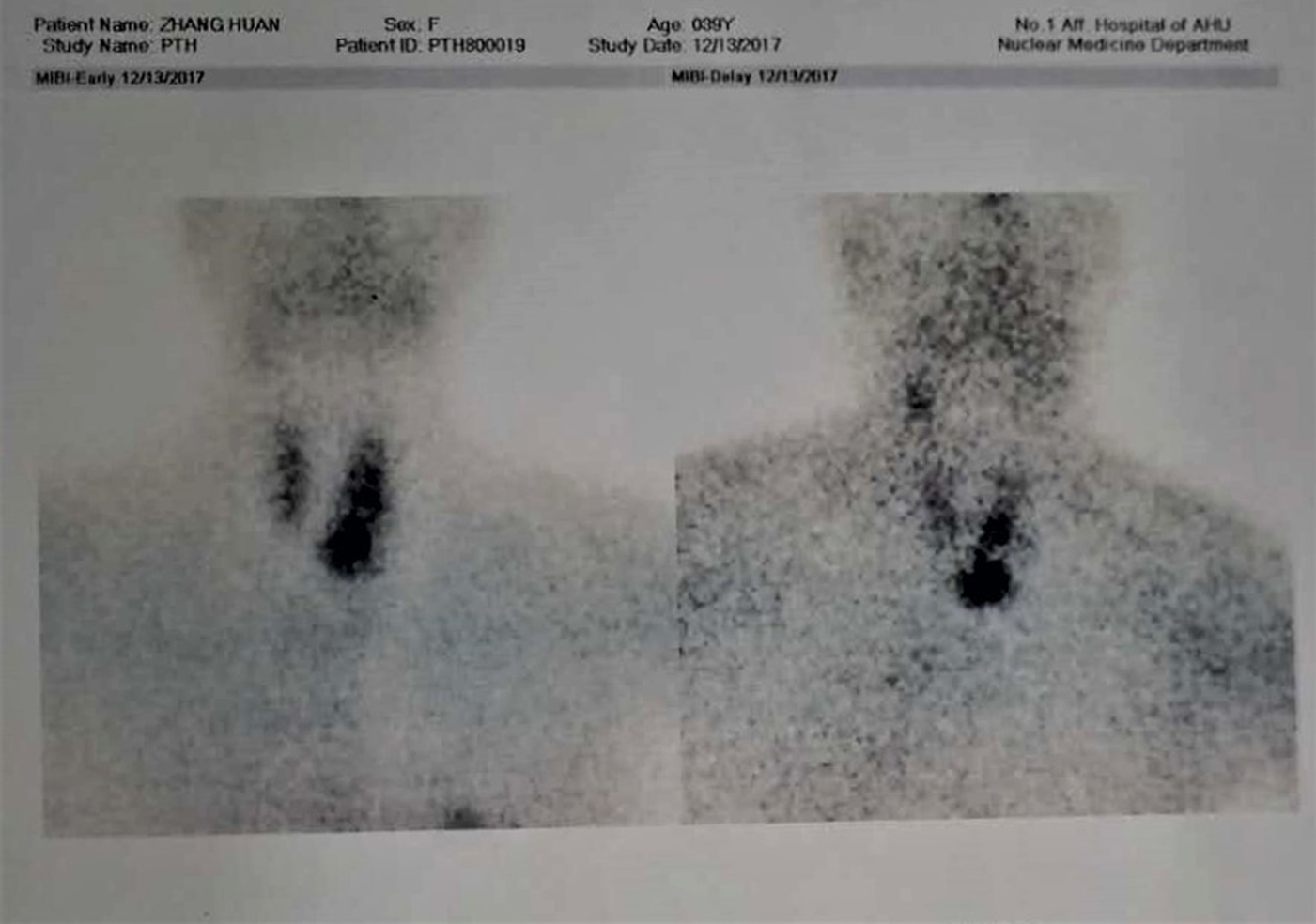
A neck ultrasound showed a right thyroid solid nodule that measured 3.3 × 2.8 mm, and fine needle aspiration revealed normal thyroid tissues. A parathyroid left inferior thyroid lobe nodule of 26 mm × 34 mm was discovered via parathyroid B- ultrasound and enhanced computed tomography scan. A 99 mTc-MIBI parathyroid imaging study was performed and showed elevated levels of radiation in the site of left inferior thyroid, which was considered as a parathyroid adenoma (Figure 1). Bone X-ray examination of the skull and whole extremities revealed no osteopenia, but bone mineral density indicated lumbar L1-4 and upper femur osteopenia. Based on the above findings, the diagnosis of primary hyperparathyroidism due to parathyroid tumor was considered.
The patient was diagnosed with primary hyperparathyroidism, left parathyroid adenoma, right thyroid adenoma, right benign mammary gland tumors and meningioma.
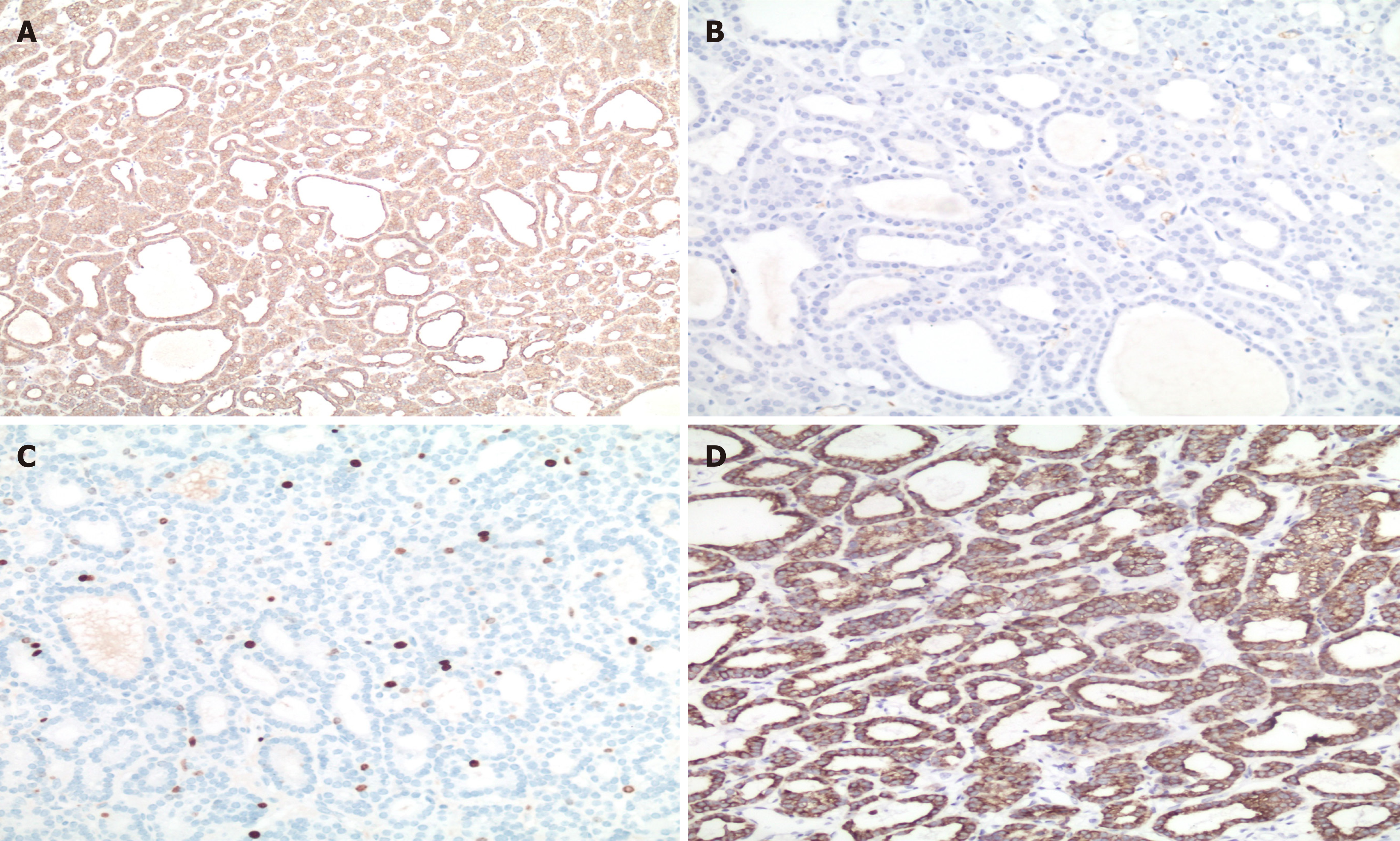
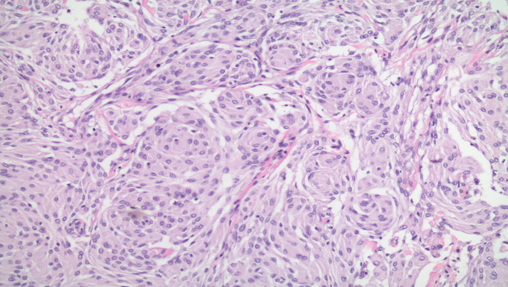
This patient received fluid replacement and calcitonin after admission. The patient underwent parathyroidectomy and the pathology revealed a parathyroid adenoma (Figure 2). The patient had undergone resection of several tumors previously, however, we only obtained the pathological picture of a meningioma (Figure 3).
The serum calcium after surgery was normal. She had recovered well after para-thyroid surgery, and her post-operative course was uneventful. Her thyroid adenoma had received routine follow-up. After myomectomy, she had not followed up recently by B-ultrasonography. In March this year, the meningioma recurred again and the patient was hospitalized.
Hyperparathyroidism involves a variety of clinical manifestations[1]. Skeletal symptoms mainly manifested as bone pain in the early stage[2]. In the late stage, skeletal malformations and pathologic fracture gradually appear, and patients even develop skull brown tumors of hyperparathyroidism[3]. This case mainly presented with digestive system manifestations. The disorders of calcium and phosphorus metabolism occurred due to elevated PTH in this patient with parathyroid adenoma. After admission to hospital, diagnosis of hyperparathyroidism was established. As is well known, the most common cause of primary hyperparathyroidism is parathyroid adenoma (80%-85%)[4]. The parathyroid tumor was found by B-ultrasound, enhanced CT scan and 99 mTc-MIBI imaging. After treatment for decreased serum calcium, this patient was switched to surgery. Her serum calcium became normal after surgical treatment.
Multiple endocrine neoplasia 1 syndrome is an autosomal dominant disorder and includes parathyroid adenoma, pituitary tumor, gastrinoma, prolactinoma, insulinoma, bronchial carcinoid, and other endocrine tumors[5]. This endocrine tumor could occur in multiple organs and systems with both benign and malignant types[6]. The neoplasms derived from the parathyroid are usually benign, while many entero-pancreatic neuroendocrine tumors and foregut carcinoid tumors are malignant. Our patient had five tumors: Left parathyroid adenoma, right thyroid adenoma, uterine fibroids, right benign mammary gland tumors, and meningoma. The patient developed five tumors successively, is it a coincidence or is there a correlation among those five tumors?
Grinblat et al[7] reported a case in a depressed woman with both meningioma and parathyroid adenoma who attempted to commit suicide. In that case, the relationship between the parathyroid adenoma and meningioma was not clarified. Unlike that case, our patient was initially found to have a meningioma and had no functional endocrine gland tumors. The thyroid adenoma was confirmed histologically by right thyroid fine needle aspiration. Patients suffering from thyroid adenoma and concurrent parathyroid adenoma are rare, with most cases in the literature reporting concurrent hot thyroid nodule and primary hyperparathyroidism[8]. Since parathyroid adenoma can be located within the thyroid, it is particularly important to differentiate between thyroid adenoma and parathyroid adenoma.
Diseases of breast, uterus, and thyroid are common in women. Spinos et al[9] reported that women with uterine fibroids had an increased incidence of thyroid nodules and fibroadenomas of the breast. In addition, some researchers reported that thyroid nodules are associated with uterine fibroids and that estrogen might have a key role in occurrence of both uterine fibroids and thyroid nodules[10]. Audisio et al[11] also maintained that estrogen might be related to the occurrence of fibroadenoma growth. During hospitalization, our patient had a normal level of estrogen, but unfortunately, we were unable to acquire her estrogen data during her entire disease course. Whether estrogen played a role in those tumors remains uncertain.
This case reported a middle-aged female who was found with multi-organ benign tumors. After surgical excision of some of these tumors, the patient recovered well. Of these tumors, the parathyroid adenoma was a hormone-secreting adenoma, and the underlying mechanisms may be revealed in future studies.
Manuscript source: Unsolicited manuscript
Specialty type: Medicine, Research and Experimental
Country of origin: China
Peer-review report classification
Grade A (Excellent): 0
Grade B (Very good): B
Grade C (Good): 0
Grade D (Fair): 0
Grade E (Poor): 0
P-Reviewer: Isik A S-Editor: Dou Y L-Editor: Filipodia E-Editor: Xing YX
| 1. | Bhansali A, Masoodi SR, Reddy KS, Behera A, das Radotra B, Mittal BR, Katariya RN, Dash RJ. Primary hyperparathyroidism in north India: a description of 52 cases. Ann Saudi Med. 2005;25:29-35. [PubMed] [DOI] [Cited in This Article: ] [Cited by in Crossref: 46] [Cited by in F6Publishing: 55] [Article Influence: 2.9] [Reference Citation Analysis (0)] |
| 2. | Udén P, Chan A, Duh QY, Siperstein A, Clark OH. Primary hyperparathyroidism in younger and older patients: symptoms and outcome of surgery. World J Surg. 1992;16:791-7; discussion 798. [PubMed] [DOI] [Cited in This Article: ] [Cited by in Crossref: 78] [Cited by in F6Publishing: 79] [Article Influence: 2.5] [Reference Citation Analysis (0)] |
| 3. | Bohdanowicz-Pawlak A, Szymczak J, Jakubowska J, Jedrzejuk D, Pawlak A, Lukienczuk T, Bolanowski M. Parathyroid adenoma diagnosed on the basis of a giant cell tumor of parieto-occipital region and multifocal bone injuries. Neuro Endocrinol Lett. 2013;34:610-614. [PubMed] [Cited in This Article: ] |
| 4. | Prasad ML, Khan A. Tumors of Parathyroid Gland. Surgical Pathology of Endocrine and Neuroendocrine Tumors. New York: Humana Press 2009; 99-110. [Cited in This Article: ] |
| 5. | Chandrasekharappa SC, Guru SC, Manickam P, Olufemi SE, Collins FS, Emmert-Buck MR, Debelenko LV, Zhuang Z, Lubensky IA, Liotta LA, Crabtree JS, Wang Y, Roe BA, Weisemann J, Boguski MS, Agarwal SK, Kester MB, Kim YS, Heppner C, Dong Q, Spiegel AM, Burns AL, Marx SJ. Positional cloning of the gene for multiple endocrine neoplasia-type 1. Science. 1997;276:404-407. [PubMed] [DOI] [Cited in This Article: ] [Cited by in Crossref: 1389] [Cited by in F6Publishing: 1203] [Article Influence: 44.6] [Reference Citation Analysis (0)] |
| 6. | Marx SJ, Agarwal SK, Kester MB, Heppner C, Kim YS, Skarulis MC, James LA, Goldsmith PK, Saggar SK, Park SY, Spiegel AM, Burns AL, Debelenko LV, Zhuang Z, Lubensky IA, Liotta LA, Emmert-Buck MR, Guru SC, Manickam P, Crabtree J, Erdos MR, Collins FS, Chandrasekharappa SC. Multiple endocrine neoplasia type 1: clinical and genetic features of the hereditary endocrine neoplasias. Recent Prog Horm Res. 1999;54:397-438; discussion 438-9. [PubMed] [DOI] [Cited in This Article: ] [Cited by in Crossref: 24] [Cited by in F6Publishing: 25] [Article Influence: 1.0] [Reference Citation Analysis (0)] |
| 7. | Grinblat J, Seidenstein B, Lerman P, Lewitus Z. Meningioma associated with parathyroid adenoma. Am J Med Sci. 1976;272:327-330. [PubMed] [DOI] [Cited in This Article: ] [Cited by in Crossref: 3] [Cited by in F6Publishing: 3] [Article Influence: 0.1] [Reference Citation Analysis (0)] |
| 8. | Sardiwalla II, Mokhtari A, Sardiwalla Y. Parathyroid adenoma with concurrent toxic thyroid adenoma: a rare combination. S Afr J Surg. 2017;55:41-44. [PubMed] [Cited in This Article: ] |
| 9. | Spinos N, Terzis G, Crysanthopoulou A, Adonakis G, Markou KB, Vervita V, Koukouras D, Tsapanos V, Decavalas G, Kourounis G, Georgopoulos NA. Increased frequency of thyroid nodules and breast fibroadenomas in women with uterine fibroids. Thyroid. 2007;17:1257-1259. [PubMed] [DOI] [Cited in This Article: ] [Cited by in Crossref: 19] [Cited by in F6Publishing: 19] [Article Influence: 1.1] [Reference Citation Analysis (0)] |
| 10. | Kim MH, Park YR, Lim DJ, Yoon KH, Kang MI, Cha BY, Lee KW, Son HY. The relationship between thyroid nodules and uterine fibroids. Endocr J. 2010;57:615-621. [PubMed] [DOI] [Cited in This Article: ] [Cited by in Crossref: 24] [Cited by in F6Publishing: 24] [Article Influence: 1.7] [Reference Citation Analysis (0)] |
| 11. | Audisio T, Crespo-Roca F, Giraudo P, Ramallo R. Fibroadenoma of the vulva--simultaneous with breast fibroadenomas and uterine myoma. J Low Genit Tract Dis. 2011;15:75-79. [PubMed] [DOI] [Cited in This Article: ] [Cited by in Crossref: 6] [Cited by in F6Publishing: 6] [Article Influence: 0.5] [Reference Citation Analysis (0)] |


