Copyright
©The Author(s) 2019.
World J Clin Cases. Sep 6, 2019; 7(17): 2644-2651
Published online Sep 6, 2019. doi: 10.12998/wjcc.v7.i17.2644
Published online Sep 6, 2019. doi: 10.12998/wjcc.v7.i17.2644
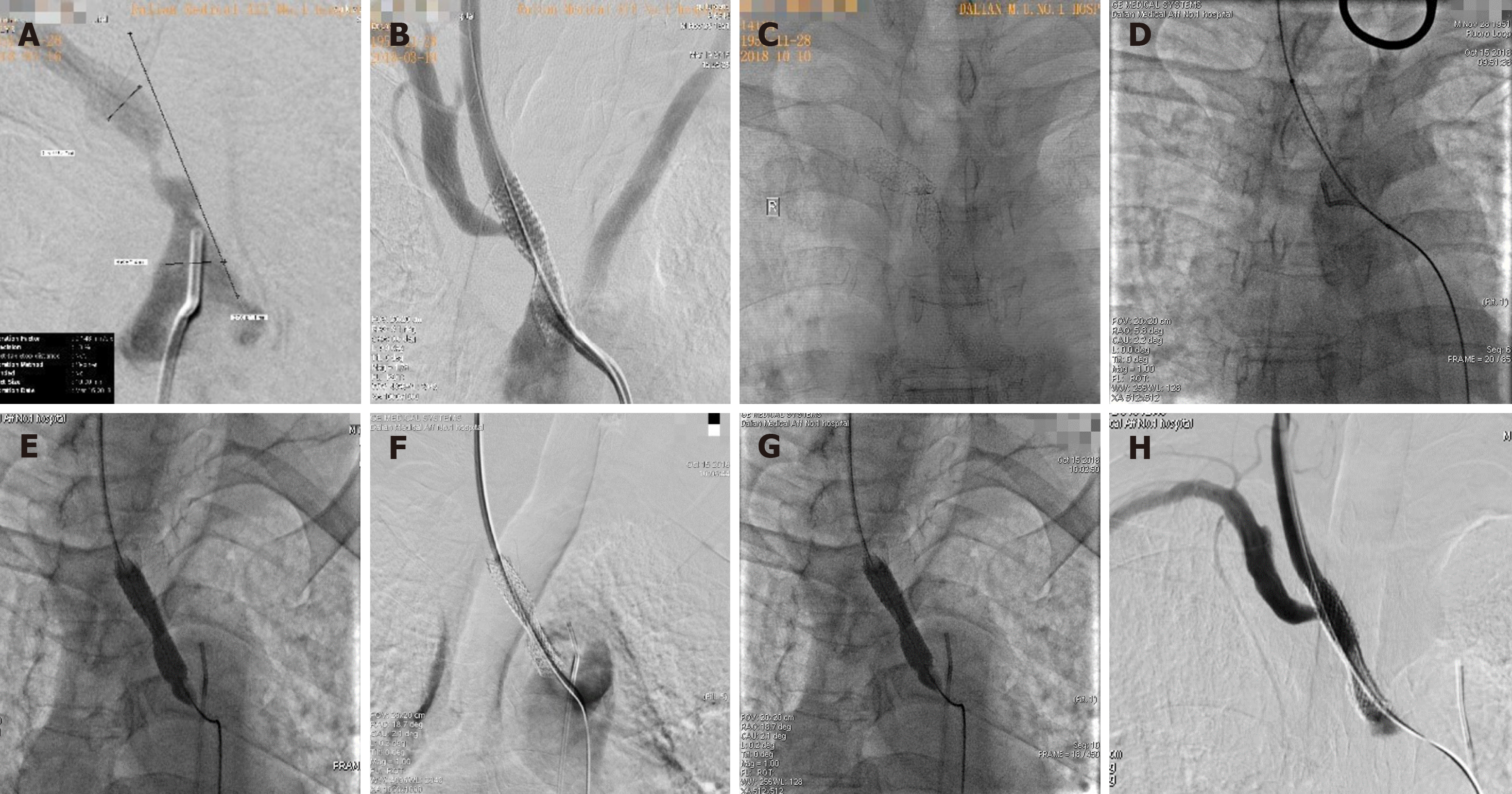
Figure 1 Cerebral angiography.
A: Cerebral angiography showing severe stenosis at the beginning of the brachiocephalic artery; B: During the first procedure, the stent was placed in the brachial artery. Cerebral angiography showed a satisfactory stent position and shape, and the blood flow velocity returned to normal; C: During the second hospitalization, cerebral angiography showed severe stenosis in the brachiocephalic artery stent; D: A carotid approach was used to smoothly pass the guidewire through the stenosis section; E: An Evercross (6 mm × 40 mm) balloon was used to expand the stenosis section; F: A Wallstent (9 mm × 30 mm) stent was placed in the stenosis, and residual stenosis was observed in the stent; G: An Evercross (8 mm × 40 mm) balloon was used to expand the residual stenosis in the stent; H: The stent was opened satisfactorily.
- Citation: Xu F, Wang F, Liu YS. Brachiocephalic artery stenting through the carotid artery: A case report and review of the literature. World J Clin Cases 2019; 7(17): 2644-2651
- URL: https://www.wjgnet.com/2307-8960/full/v7/i17/2644.htm
- DOI: https://dx.doi.org/10.12998/wjcc.v7.i17.2644


