Copyright
©The Author(s) 2019.
World J Clin Cases. Aug 6, 2019; 7(15): 2049-2057
Published online Aug 6, 2019. doi: 10.12998/wjcc.v7.i15.2049
Published online Aug 6, 2019. doi: 10.12998/wjcc.v7.i15.2049
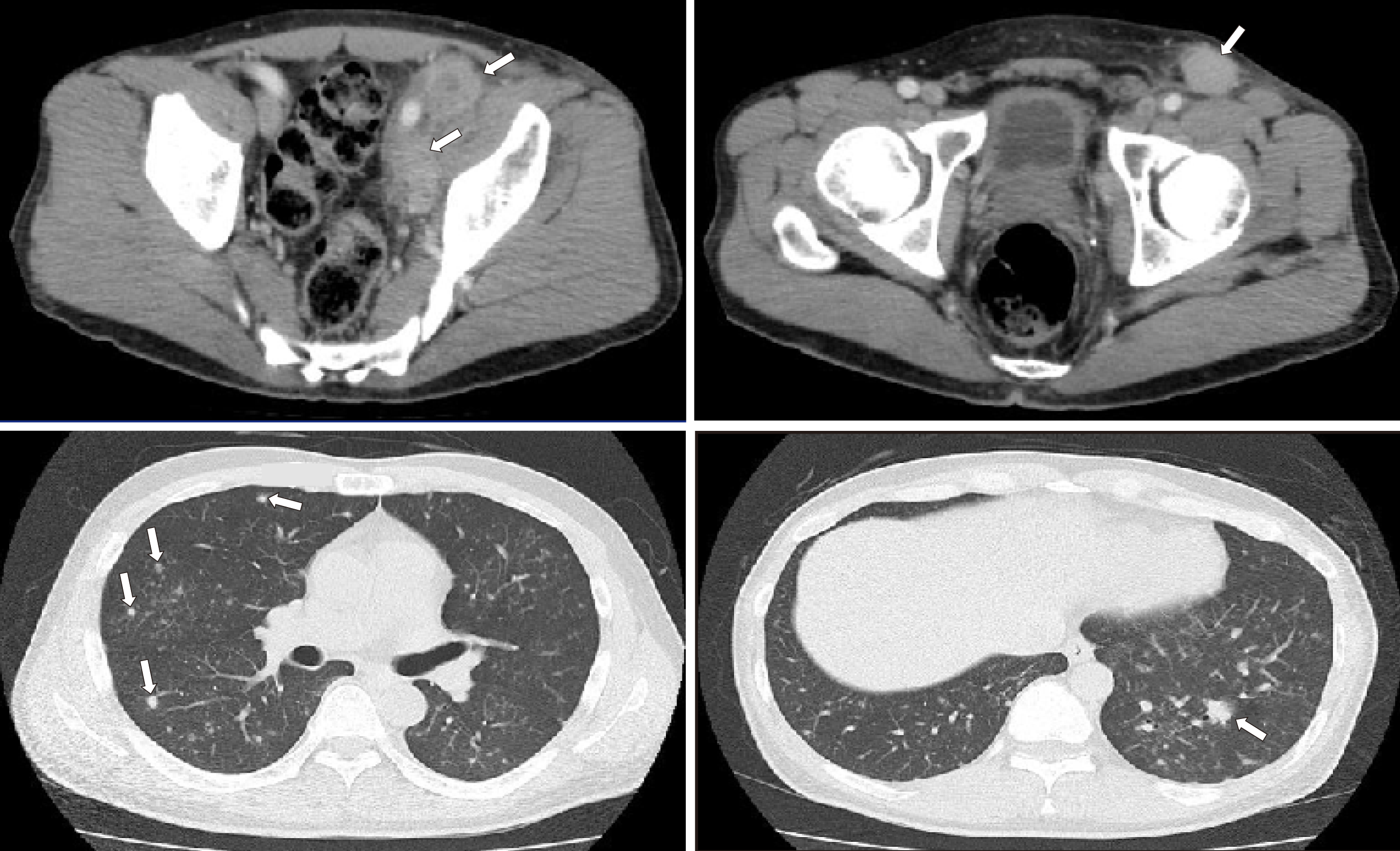
Figure 1 Computed tomography images.
(Upper row) Pelvic computed tomography scan demonstrated multiple lymph node lesions (arrows) continuing to the left common iliac artery (left)/left inguinal region (right). (Lower row) Scattered nodular lesions were found in both lung fields (arrows).
- Citation: Saito M, Izumiyama K, Ogasawara R, Mori A, Kondo T, Tanaka M, Morioka M, Miyashita K, Tanino M. ALK-positive anaplastic large cell lymphoma presenting multiple lymphomatous polyposis: A case report and literature review. World J Clin Cases 2019; 7(15): 2049-2057
- URL: https://www.wjgnet.com/2307-8960/full/v7/i15/2049.htm
- DOI: https://dx.doi.org/10.12998/wjcc.v7.i15.2049


