Copyright
©The Author(s) 2022.
World J Clin Cases. Nov 26, 2022; 10(33): 12313-12318
Published online Nov 26, 2022. doi: 10.12998/wjcc.v10.i33.12313
Published online Nov 26, 2022. doi: 10.12998/wjcc.v10.i33.12313
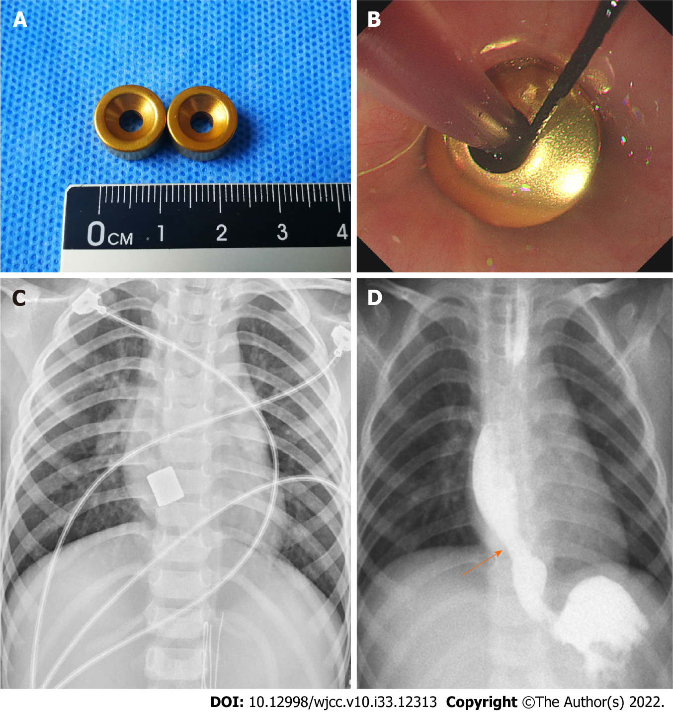
Figure 2 Magnetic compression stricturoplasty.
A: Magnets prepared for the operation; B: Magnets were placed under endoscopy and fluoroscopy guidance; C: Intraoperative chest radiograph confirming that the magnets were in good alignment; D: Magnets were removed and radiographic examination of the patient after magnetic compression stricturoplasty showed a patent esophageal lumen with an inner diameter of 8.5 mm at the stenosis site on day 14 (arrow).
- Citation: Liu SQ, Lv Y, Luo RX. Endoscopic magnetic compression stricturoplasty for congenital esophageal stenosis: A case report. World J Clin Cases 2022; 10(33): 12313-12318
- URL: https://www.wjgnet.com/2307-8960/full/v10/i33/12313.htm
- DOI: https://dx.doi.org/10.12998/wjcc.v10.i33.12313


