Published online Nov 16, 2022. doi: 10.12998/wjcc.v10.i32.11936
Peer-review started: June 29, 2022
First decision: September 5, 2022
Revised: September 7, 2022
Accepted: October 11, 2022
Article in press: October 11, 2022
Published online: November 16, 2022
Symptomatic cervical facet cysts are relatively rare compared to those in the lumbar region. These cysts are usually located in the 7th cervical and 1st thoracic vertebral (C7/T1) area, and surgical excision is performed in most cases. However, facet cysts are associated with degenerative conditions, and elderly patients are often ineligible for surgical procedures. Cervical interlaminar epidural block has been used in patients with cervical radiating symptoms and achieved good results. Therefore, cervical interlaminar epidural block may be the first-choice treatment for symptomatic cervical facet cysts.
A 70-year-old man complained of a tingling sensation in the left hand, focused on the 4th and 5th fingers, for 1 year, and posterior neck pain for over 5 mo. The patient’s numeric rating scale (NRS) score was 5/10. The patient was diagnosed with symptomatic cervical facet cyst at the left C7/T1 facet joint. Fluoroscopy-guided cervical interlaminar epidural block at the C7/T1 level with 20 mg triamcinolone and 5 mL of 0.5% lidocaine was administered. The patient's symptoms improved immediately after the block, with an NRS score of 3 points. After 3 mo, his left posterior neck pain and tingling along the left 8th cervical dermatome were relieved, with an NRS score of 2.
A cervical interlaminar epidural block is a good alternative for managing symptomatic cervical facet cysts.
Core Tip: Intraspinal facet cysts are usually asymptomatic and incidentally identified. However, they could be symptomatic and interfere with the patient’s quality of life. Symptomatic cervical facet cysts are relatively rare compared to those in the lumbar region. Guidelines for the management of cervical facet cysts have not been well established. Several patients reported in the existing literature underwent surgery for symptomatic cervical facet cysts, but there are situations where surgery may not be possible, such as an underlying disorder or refusal of the patient. In such cases, a cervical interlaminar epidural block can be a good alternative.
- Citation: Hwang SM, Lee MK, Kim S. Management of symptomatic cervical facet cyst with cervical interlaminar epidural block: A case report. World J Clin Cases 2022; 10(32): 11936-11941
- URL: https://www.wjgnet.com/2307-8960/full/v10/i32/11936.htm
- DOI: https://dx.doi.org/10.12998/wjcc.v10.i32.11936
A facet cyst is defined as extrusion of the synovium through a capsular defect, resulting from an unstable or degenerate synovial joint. Symptomatic intraspinal facet cysts are typically located on the lumbar spine. Facet cysts of the cervical spine are rarely reported. Despite several decades of research, the etiology of intraspinal facet cysts remains unclear. Extrusion of synovial fluid through defects and tears of the facet capsule, degeneration of myxoid, and cyst formation from collagen tissue have been proposed as potential etiologies[1]. Moreover, synovial extrusion can be aggravated by hypermobility or inflammatory factors resulting in radiating symptoms. The most common treatment for symptomatic cervical facet cysts is the surgical removal of the cyst through hemilaminectomy or bilateral laminectomy, with or without instrumented fusion[2]. Non-surgical management of cervical facet cysts is rarely applicable. Here, we present a rare case of symptomatic cervical facet cyst managed with cervical interlaminar epidural block.
A 70-year-old man visited our pain clinic with posterior neck pain, tingling sensation, and numbness along the left 8th cervical (C8) vertebral dermatome.
A tingling sensation and numbness in the left hand, localized to the 4th and 5th finger, started a year earlier. The patient subsequently complained of posterior neck pain for more than 5 mo.
Before visiting our clinic, he was treated with oral pregabalin (150 mg/d), duloxetine (30 mg/d), and tramadol (100 mg/d) for 2 wk in the neurosurgery clinic of our hospital. However, the patient did not respond to this treatment. We used the numeric rating scale (NRS) to assess the severity of pain, and the patient's NRS score was 5 out of 10. His symptoms worsened when he was lying on his left side.
The patient had hypertension and a history of angina pectoris but no history of cervical trauma. The patient had no specific family history.
Physical examination revealed sensory impairment in the left C8 dermatome, but motor functions were intact. Spurling's test showed a positive result on the left side. The deep tendon reflexes were normal.
Laboratory examinations did not reveal any abnormalities.
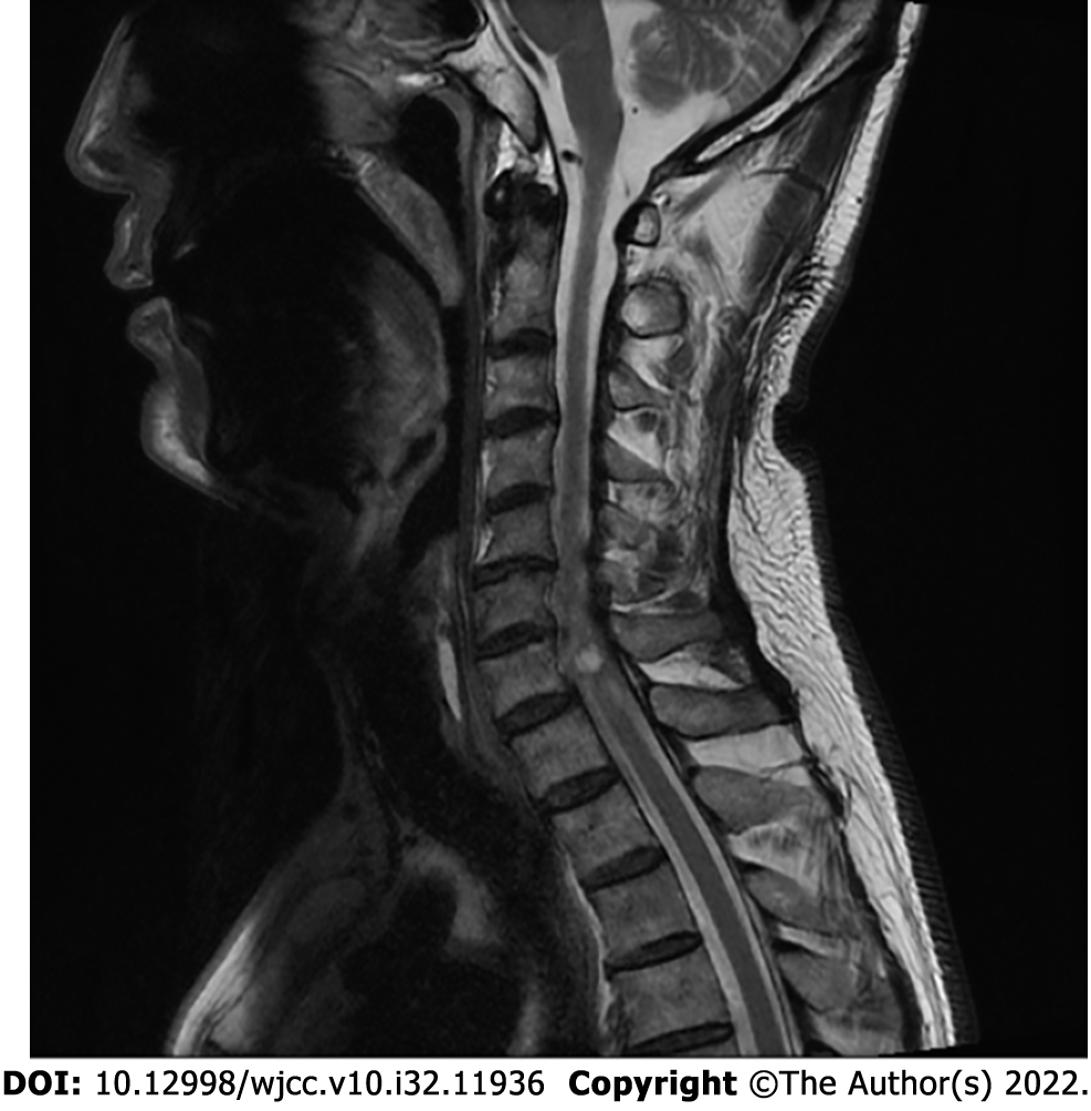
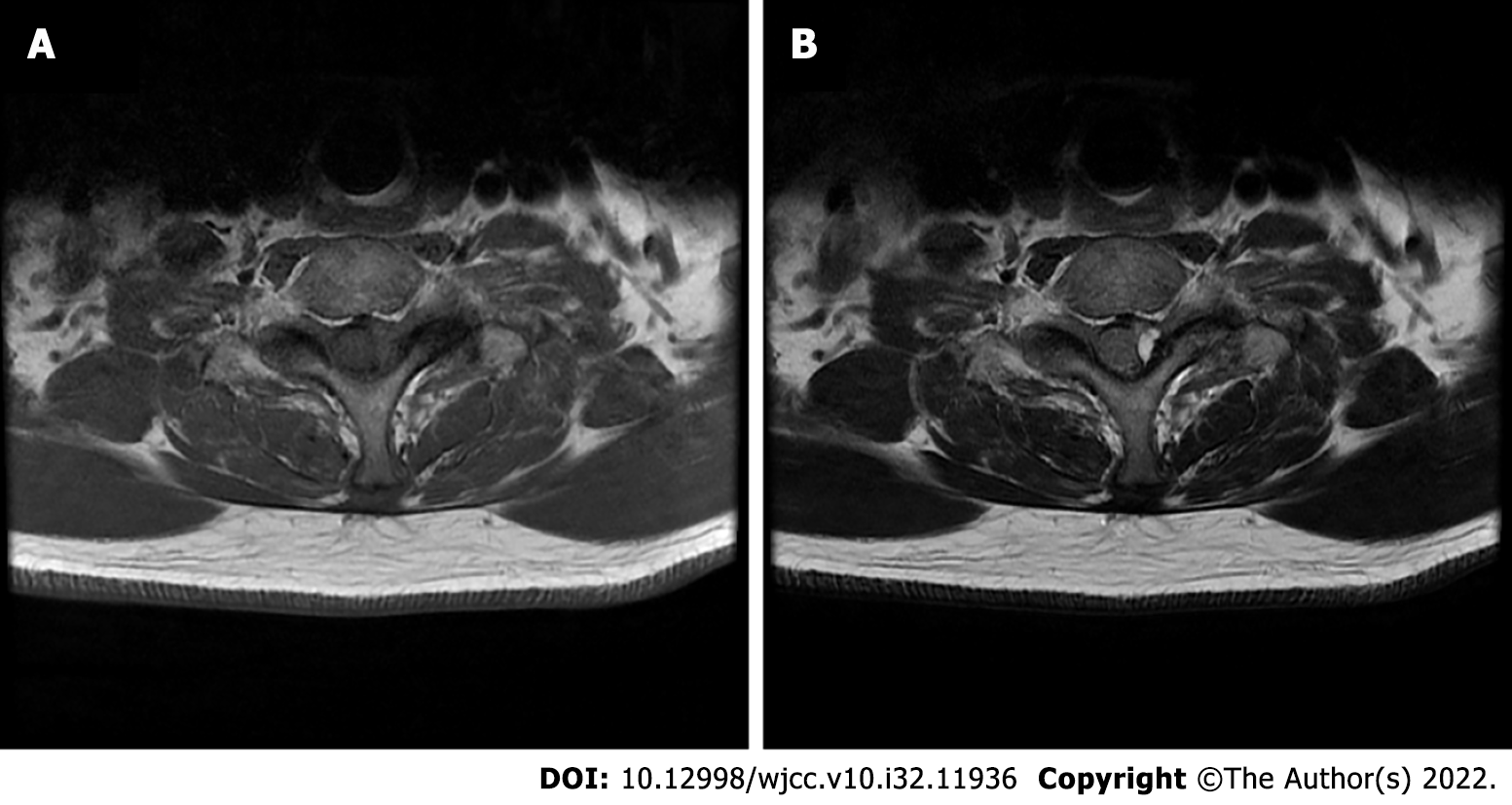
Magnetic resonance imaging (MRI) of the cervical spine revealed a 6 mm × 6 mm × 10 mm intradural extramedullary tumor cyst on the left posterolateral aspect of the 7th cervical and 1st thoracic vertebral (C7/T1) facet joint (Figure 1). The cyst showed a low signal intensity on T1-weighted images. On T2-weighted images, high signal intensity in the center and low signal intensity around the rim were observed (Figure 2).
The patient was diagnosed with symptomatic cervical facet cyst at the left C7/T1 facet joint.
Although symptomatic cervical facet cysts are usually treated with surgical excision, the patient was worried about surgery because of his old age and cardiac condition. Therefore, we decided to perform nonsurgical nerve block. A fluoroscopy-guided cervical interlaminar epidural block was performed at the C7/T1 level using 20 mg of triamcinolone and 5 mL of 0.5% lidocaine.
The patient's symptoms improved immediately after the procedure, and the NRS score decreased to 3 points. The patient revisited the clinic for follow-up after 2 wk. His left posterior neck pain and tingling along the left C8 dermatome were relieved, with an NRS score of 2 points. The patient further reported that his symptoms had improved and he no longer required medication. There were no complications associated with the block procedure. At the 3 mo follow-up after the cervical interlaminar epidural block, the patient's pain was still rated as 2 on the NRS, although the numbness persisted. The patient was satisfied with the clinical outcome and refused follow-up MRI because of the symptomatic improvement and financial burden.
Cervical facet cysts were first described in 1985 by Cartwright et al[3]. They are often found at the C7/T1 level and are relatively uncommon compared to lumbar facet cysts[4]. Intraspinal facet cysts are usually asymptomatic and incidentally identified. However, they could be symptomatic and interfere with the patient’s quality of life. The clinical presentation of symptomatic intraspinal facet cysts includes radicular pain (84%), back pain before radicular pain (50%-93%), neurologic claudication (10%-44%), sensory deficits (10%-43%), motor deficits (20%-27%), and symptoms consistent with cauda equina syndrome[5]. Our patient also experienced numbness and tingling sensation along the left C8 dermatome and left posterior neck pain.
MRI is the most sensitive imaging modality for diagnosing cervical facet cysts. On MRI, these cysts appeared as well-circumscribed extradural cystic lesions close to the facet joints. These lesions have a low signal intensity on T1-weighted images and high signal intensity on T2-weighted images. Facet cysts show a typical pattern of high signal intensity at the center and low signal intensity around the rim[6]. We observed an image of the cyst at the left C7/T1 level on the T2-weighted MRI.
Guidelines for the management of cervical facet cysts have not been well established. Patients with significant neurological deficits, motor weakness, intractable pain, or multiple synovial cysts should undergo surgical treatment to achieve the best long-term outcome[5]. According to previous reports, surgical removal of symptomatic cervical facet cysts through hemilaminectomy and bilateral laminectomy, with or without instrumented fusion, is usually performed[2,4]. Although surgical treatment is generally successful, with complete removal of the cyst, from the patient’s point of view, it can be a burdensome choice as first-line treatment. In addition, the possibility of complications related to surgery and anesthesia should be considered. Therefore, nonsurgical management can be an alternative to reduce the surgical burden.
Regarding symptomatic lumbar facet cysts, there have been several reports of conservative management such as percutaneous cyst aspiration and percutaneous steroid injection. Unger et al[7] reported a case of successful management of symptomatic L4/5 facet cysts by using L4/5 and L5/S1 transforaminal epidural steroid injections. Slipman et al[8] further evaluated nonsurgical managements, such as therapeutic selective nerve root block, intraarticular zygapophyseal joint corticosteroid injection, and cyst puncture for symptomatic lumbar facet cysts. They reported that with follow-up for an average of 1.4 years after the termination of management, one-third of the patients (28.6%) were found to have an excellent outcome. Lee et al[9] successfully managed a symptomatic L5/S1 facet joint cyst and left S1 radiculopathy using percutaneous treatment with steroid injection and distension of the cyst. Jin et al[10] reported successful removal of an L4/5 facet cyst by mechanical rupture under epiduroscopic guidance.
Although there have been several reports of conservative management of lumbar facet cysts, studies of cervical facet cysts are relatively rare. Kostanian et al[11] reported the successful management of symptomatic cervical facet cysts at C6/7 by cyst aspiration under computed tomography guidance combined with epidural injection of a local anesthetic and steroid. Tarukado et al[12] suggested the possibility of spontaneous regression of a symptomatic C4/5 facet cyst; thus, conservative management might be indicated for symptomatic cervical facet cysts, especially in cases of radiculopathy of the middle cervical spine.
We believe that symptoms caused by cervical facet cysts can be managed similarly to those caused by cervical herniated nucleus pulposus or cervical spinal stenosis. Therefore, a cervical interlaminar epidural block could be the first choice for the management of symptomatic cervical facet cysts. If cervical interlaminar epidural block fails, other conservative options, including image-guided aspiration of the cyst, epiduroscopic removal, or surgery, can be considered. Therefore, we performed a fluoroscopy-guided cervical interlaminar epidural block at the C7/T1 level using 20 mg of triamcinolone and 5 mL of 0.5% lidocaine as the first treatment choice to relieve symptoms. Mixtures of local anesthetics and corticosteroids have been used for epidural block. The rationale for injecting local anesthetics is to block sensory signals. The effectiveness of local anesthetics for chronic pain is based on their anti-inflammatory actions and the alteration of multiple pathophysiological mechanisms. Moreover, corticosteroids in epidural block have been used to reduce inflammation either by inhibiting the synthesis or release of a number of proinflammatory substances or by causing a reversible local anesthetic effect[13]. The patient’s symptoms were sufficiently reduced after the procedure. The patient refused follow-up cervical MRI because of symptomatic improvement and financial burden. Therefore, it was not possible to determine whether the cervical facet cyst regressed spontaneously as in a previous report[12], decreased in size compared to the previous size, or whether the cervical facet cyst persisted; however, the patient’s symptoms were improved after the cervical interlaminar epidural block.
Several patients reported in the existing literature underwent surgery for symptomatic cervical facet cysts, but there are situations where surgery may not be possible, such as an underlying disorder or refusal of the patient. In such cases, a cervical interlaminar epidural block can be a good alternative.
In conclusion, there are various options for the management of symptomatic cervical facet cysts. However, standard management guidelines have not yet been established. Therefore, we need to consider the value of cervical interlaminar epidural block as an effective conservative approach, and more attention should be paid to conservative management to improve the clinical outcomes in various patient conditions.
Provenance and peer review: Unsolicited article; Externally peer reviewed.
Peer-review model: Single blind
Specialty type: Medicine, research and experimental
Country/Territory of origin: South Korea
Peer-review report’s scientific quality classification
Grade A (Excellent): 0
Grade B (Very good): B, B
Grade C (Good): 0
Grade D (Fair): 0
Grade E (Poor): 0
P-Reviewer: Gao L, China; Naem AA, Syria S-Editor: Liu GL L-Editor: A P-Editor: Liu GL
| 1. | Phan K, Mobbs RJ. A rare case of cervical facet joint and synovial cyst at C5/C6. J Clin Neurosci. 2016;29:191-194. [PubMed] [DOI] [Cited in This Article: ] [Cited by in Crossref: 1] [Cited by in F6Publishing: 1] [Article Influence: 0.1] [Reference Citation Analysis (62)] |
| 2. | Uschold T, Panchmatia J, Fusco DJ, Abla AA, Porter RW, Theodore N. Subaxial cervical juxtafacet cysts: single institution surgical experience and literature review. Acta Neurochir (Wien). 2013;155:299-308. [PubMed] [DOI] [Cited in This Article: ] [Cited by in Crossref: 8] [Cited by in F6Publishing: 10] [Article Influence: 0.9] [Reference Citation Analysis (2)] |
| 3. | Cartwright MJ, Nehls DG, Carrion CA, Spetzler RF. Synovial cyst of a cervical facet joint: case report. Neurosurgery. 1985;16:850-852. [PubMed] [DOI] [Cited in This Article: ] [Cited by in Crossref: 65] [Cited by in F6Publishing: 65] [Article Influence: 1.7] [Reference Citation Analysis (1)] |
| 4. | Lyons MK, Birch BD, Krauss WE, Patel NP, Nottmeier EW, Boucher OK. Subaxial cervical synovial cysts: report of 35 histologically confirmed surgically treated cases and review of the literature. Spine (Phila Pa 1976). 2011;36:E1285-E1289. [PubMed] [DOI] [Cited in This Article: ] [Cited by in Crossref: 23] [Cited by in F6Publishing: 24] [Article Influence: 1.8] [Reference Citation Analysis (0)] |
| 5. | Bydon M, Papadimitriou K, Witham T, Wolinsky JP, Sciubba D, Gokaslan Z, Bydon A. Treatment of spinal synovial cysts. World Neurosurg. 2013;79:375-380. [PubMed] [DOI] [Cited in This Article: ] [Cited by in Crossref: 16] [Cited by in F6Publishing: 20] [Article Influence: 1.8] [Reference Citation Analysis (0)] |
| 6. | Schmid G, Willburger R, Jergas M, Pennekamp W, Bickert U, Köster O. [Lumbar intraspinal juxtafacet cysts: MR imaging and CT-arthrography]. Rofo. 2002;174:1247-1252. [PubMed] [DOI] [Cited in This Article: ] [Cited by in Crossref: 17] [Cited by in F6Publishing: 18] [Article Influence: 0.8] [Reference Citation Analysis (0)] |
| 7. | Unger R, Bajaj PS. Poster 410 treatment of a lumbar facet joint synovial cyst using fluoroscopic guided transforaminal epidural steroid injection: A case report. Pm & R. 4:S331-S331. [DOI] [Cited in This Article: ] [Cited by in F6Publishing: 1] [Reference Citation Analysis (0)] |
| 8. | Slipman CW, Lipetz JS, Wakeshima Y, Jackson HB. Nonsurgical treatment of zygapophyseal joint cyst-induced radicular pain. Arch Phys Med Rehabil. 2000;81:973-977. [PubMed] [DOI] [Cited in This Article: ] [Cited by in Crossref: 21] [Cited by in F6Publishing: 21] [Article Influence: 0.9] [Reference Citation Analysis (0)] |
| 9. | Lee SJ, Kim YK, Jung HS, Lim JB, Lee C. Percutaneous treatment with steroid injections and distension of facet synovial cyst-a case report. Korean J Pain. 2005;18:246-250. [DOI] [Cited in This Article: ] [Cited by in Crossref: 1] [Cited by in F6Publishing: 1] [Article Influence: 0.1] [Reference Citation Analysis (0)] |
| 10. | Jin HS, Bae JY, In CB, Choi EJ, Lee PB, Nahm FS. Epiduroscopic Removal of a Lumbar Facet Joint Cyst. Korean J Pain. 2015;28:275-279. [PubMed] [DOI] [Cited in This Article: ] [Cited by in Crossref: 8] [Cited by in F6Publishing: 8] [Article Influence: 0.9] [Reference Citation Analysis (0)] |
| 11. | Kostanian VJ, Mathews MS. CT guided aspiration of a cervical synovial cyst. Case report and technical note. Interv Neuroradiol. 2007;13:295-298. [PubMed] [DOI] [Cited in This Article: ] [Cited by in Crossref: 10] [Cited by in F6Publishing: 13] [Article Influence: 0.8] [Reference Citation Analysis (1)] |
| 12. | Tarukado K, Uemori T, Ueda S, Imamura T, Kaji K. Spontaneous regression of a subaxial cervical facet cyst. Interdiscip Neurosurg. 2021;23:100952. [DOI] [Cited in This Article: ] [Reference Citation Analysis (0)] |
| 13. | Manchikanti L, Knezevic NN, Navani A, Christo PJ, Limerick G, Calodney AK, Grider J, Harned ME, Cintron L, Gharibo CG, Shah S, Nampiaparampil DE, Candido KD, Soin A, Kaye AD, Kosanovic R, Magee TR, Beall DP, Atluri S, Gupta M, Helm Ii S, Wargo BW, Diwan S, Aydin SM, Boswell MV, Haney BW, Albers SL, Latchaw R, Abd-Elsayed A, Conn A, Hansen H, Simopoulos TT, Swicegood JR, Bryce DA, Singh V, Abdi S, Bakshi S, Buenaventura RM, Cabaret JA, Jameson J, Jha S, Kaye AM, Pasupuleti R, Rajput K, Sanapati MR, Sehgal N, Trescot AM, Racz GB, Gupta S, Sharma ML, Grami V, Parr AT, Knezevic E, Datta S, Patel KG, Tracy DH, Cordner HJ, Snook LT, Benyamin RM, Hirsch JA. Epidural Interventions in the Management of Chronic Spinal Pain: American Society of Interventional Pain Physicians (ASIPP) Comprehensive Evidence-Based Guidelines. Pain Physician. 2021;24:S27-S208. [PubMed] [Cited in This Article: ] |


