Copyright
©The Author(s) 2022.
World J Clin Cases. Aug 16, 2022; 10(23): 8384-8391
Published online Aug 16, 2022. doi: 10.12998/wjcc.v10.i23.8384
Published online Aug 16, 2022. doi: 10.12998/wjcc.v10.i23.8384
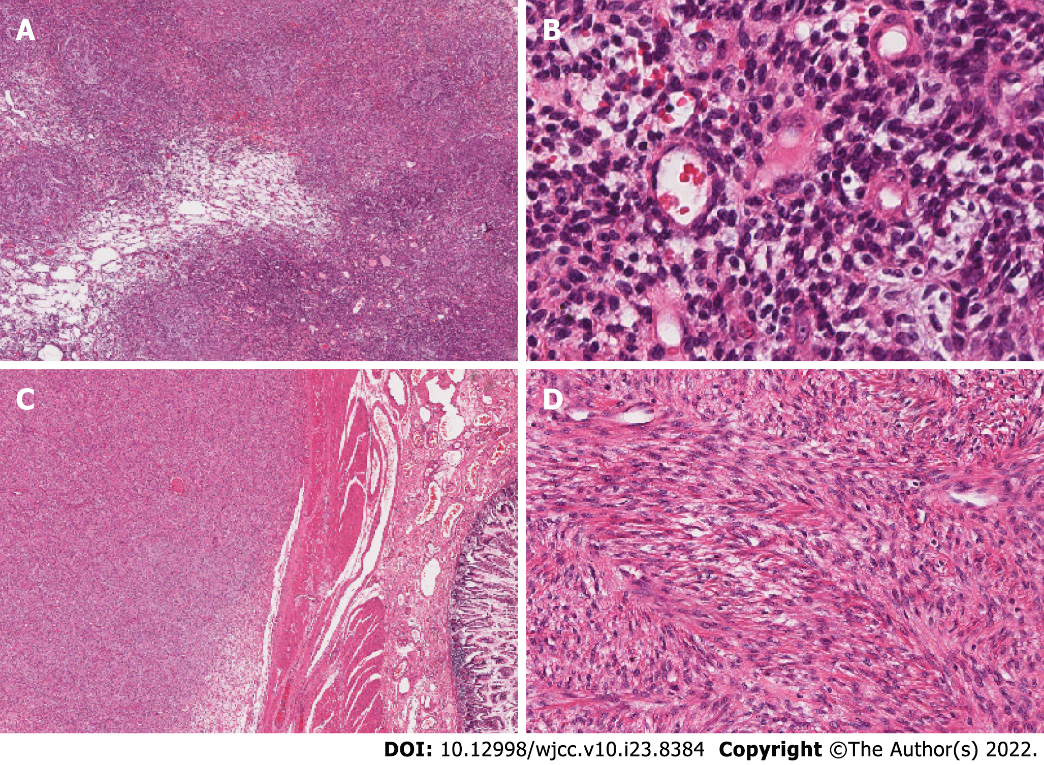
Figure 2 The right ovarian mass and the mesenteric mass in hematoxylin and eosin stain.
A: The ovarian tumor has grown as a solid sheet (Original magnification: 40 ×; scale bar: 100 μm); B: In some areas, tumors cells have grown around small blood vessels (Original magnification: 200 ×; scale bar: 100 μm); C: The mesenteric tumor exhibits local invasion of the intestinal serosa and underlying muscle (Original magnification: 40 ×; scale bar: 100 μm); D: Cells have grown in a fishbone-like arrangement (Original magnification: 100 ×; scale bar: 100 μm).
- Citation: Yu HY, Jin YL. Metastatic low-grade endometrial stromal sarcoma with variable morphologies in the ovaries and mesentery: A case report. World J Clin Cases 2022; 10(23): 8384-8391
- URL: https://www.wjgnet.com/2307-8960/full/v10/i23/8384.htm
- DOI: https://dx.doi.org/10.12998/wjcc.v10.i23.8384


