Copyright
©The Author(s) 2022.
World J Clin Cases. Jun 26, 2022; 10(18): 6298-6306
Published online Jun 26, 2022. doi: 10.12998/wjcc.v10.i18.6298
Published online Jun 26, 2022. doi: 10.12998/wjcc.v10.i18.6298
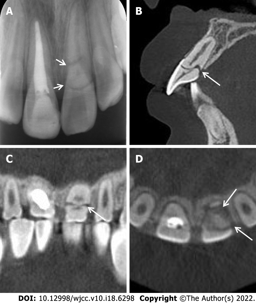
Figure 7 Periapical radiograph and cone beam computed tomography images.
A: Periapical radiograph of teeth 11 and 21 taken 9 mo after the restoration of tooth 11; B-D: Sagittal (B), coronal (C), and cross-sectional (D) cone beam computed tomography images taken 4 yr later showing sign of repair.
- Citation: Zhou ZL, Gao L, Sun SK, Li HS, Zhang CD, Kou WW, Xu Z, Wu LA. Spontaneous healing of complicated crown-root fractures in children: Two case reports. World J Clin Cases 2022; 10(18): 6298-6306
- URL: https://www.wjgnet.com/2307-8960/full/v10/i18/6298.htm
- DOI: https://dx.doi.org/10.12998/wjcc.v10.i18.6298


