Copyright
©The Author(s) 2022.
World J Clin Cases. Jun 26, 2022; 10(18): 6277-6282
Published online Jun 26, 2022. doi: 10.12998/wjcc.v10.i18.6277
Published online Jun 26, 2022. doi: 10.12998/wjcc.v10.i18.6277
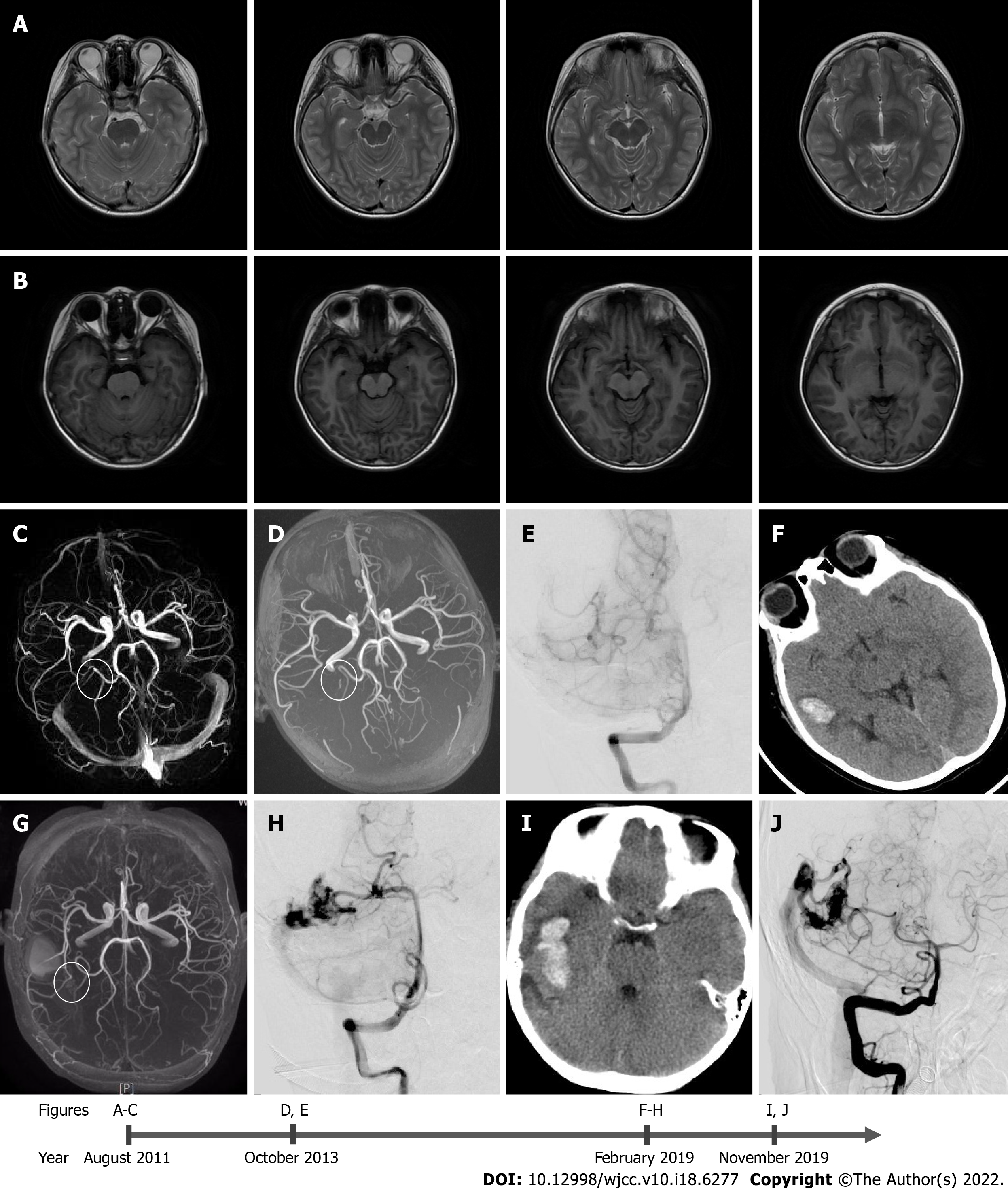
Figure 1 Diagnostic images from August 2011 to November 2019.
A-C: T1 (A) and T2 (B) weighted images of magnetic resonance imaging, and (C) magnetic resonance angiography (MRA) done in August 2011, at the age of 5 years and 2 mo. There is no evidence of vascular lesion; D and E: The results of (D) MRA and (E) the first anteroposterior digital subtraction angiography (DSA) performed in October 2013, indicating de novo arteriovenous malformation; F-H: The results of (F) the head computed tomography (CT) scan, (G) MRA, and (H) the second DSA, which were performed in February 2019; I and J: Results from (I) the post-hemorrhage non-contrast head CT scan depicting blood in the right temporal lobe and (J) the third DSA, which were performed in November 2019.
- Citation: Huang H, Wang X, Guo AN, Li W, Duan RH, Fang JH, Yin B, Li DD. De novo brain arteriovenous malformation formation and development: A case report. World J Clin Cases 2022; 10(18): 6277-6282
- URL: https://www.wjgnet.com/2307-8960/full/v10/i18/6277.htm
- DOI: https://dx.doi.org/10.12998/wjcc.v10.i18.6277


