Copyright
©The Author(s) 2022.
World J Clin Cases. Jun 26, 2022; 10(18): 6227-6233
Published online Jun 26, 2022. doi: 10.12998/wjcc.v10.i18.6227
Published online Jun 26, 2022. doi: 10.12998/wjcc.v10.i18.6227
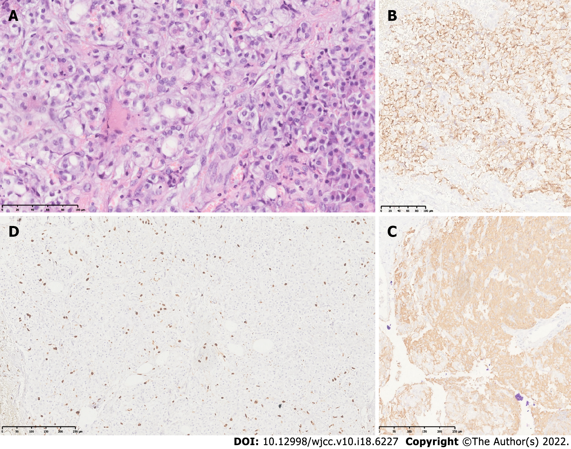
Figure 2 Histology and immunohistochemistry of the tumor.
A: Hematoxylin and eosin photomicrograph (40 ×) showing neoplastic cells presenting uniformly round nuclei with granular chromatin (salt and pepper image), typical of neuroendocrine cells with extensive eosinophilic cytoplasm; B and C: Photomicrographs of immunohistochemical staining for chromogranin (B) and synaptophysin (C) (10 ×). Neoplastic cells show strong immunopositivity for both markers in the cytoplasm, which corroborates the neuroendocrine lineage of the neoplasia; D: Immunohistochemical staining for Ki67 (cell proliferation index). Strong nuclear immunopositivity is seen in approximately 4% of neoplastic cells. The tumor was classified as grade 2, according to the 2019 WHO classification.
- Citation: Lobaton-Ginsberg M, Sotelo-González P, Ramirez-Renteria C, Juárez-Aguilar FG, Ferreira-Hermosillo A. Insulinoma after sleeve gastrectomy: A case report. World J Clin Cases 2022; 10(18): 6227-6233
- URL: https://www.wjgnet.com/2307-8960/full/v10/i18/6227.htm
- DOI: https://dx.doi.org/10.12998/wjcc.v10.i18.6227


