Copyright
©The Author(s) 2022.
World J Clin Cases. May 6, 2022; 10(13): 4214-4219
Published online May 6, 2022. doi: 10.12998/wjcc.v10.i13.4214
Published online May 6, 2022. doi: 10.12998/wjcc.v10.i13.4214
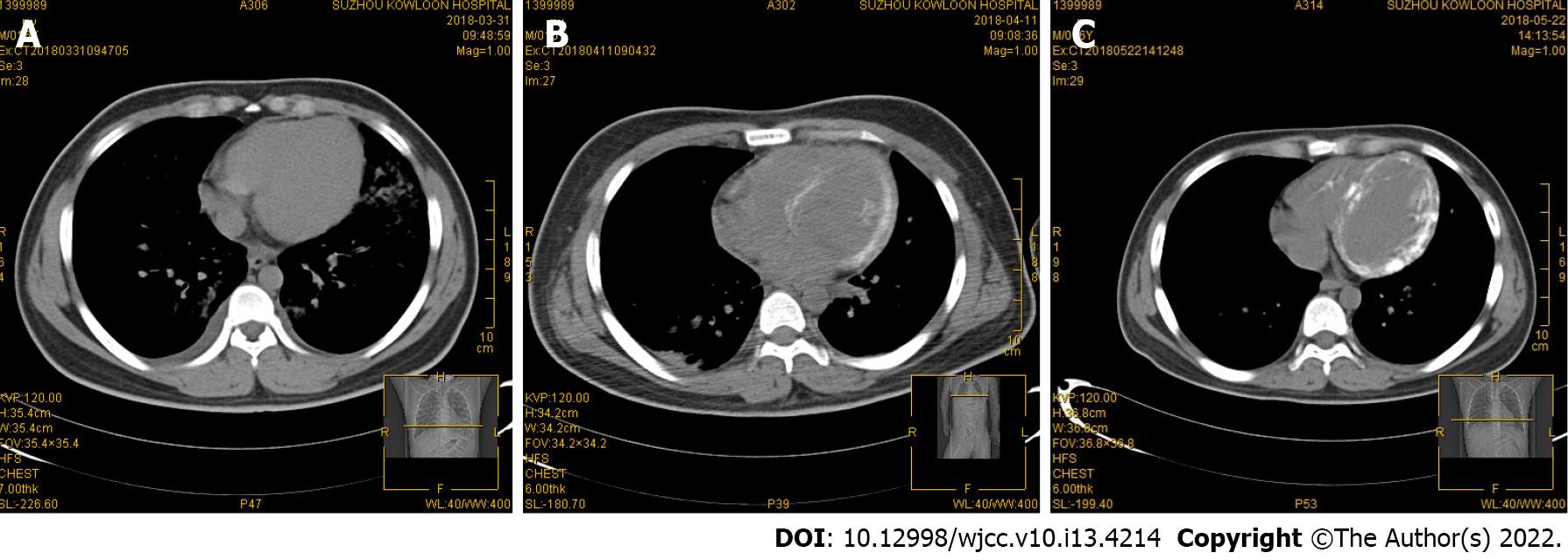
Figure 1 Chest computed tomography findings.
A: Chest computed tomography (CT) on the day of admission showed no morphological abnormalities in the heart; B: After 10 d, chest CT showed an increase in left ventricular wall density; C: 30 d later, CT showed obvious myocardial calcification in the left ventricle.
- Citation: Sui ML, Wu CJ, Yang YD, Xia DM, Xu TJ, Tang WB. Extensive myocardial calcification in critically ill patients receiving extracorporeal membrane oxygenation: A case report. World J Clin Cases 2022; 10(13): 4214-4219
- URL: https://www.wjgnet.com/2307-8960/full/v10/i13/4214.htm
- DOI: https://dx.doi.org/10.12998/wjcc.v10.i13.4214


