Copyright
©2014 Baishideng Publishing Group Inc.
World J Nephrol. Nov 6, 2014; 3(4): 122-142
Published online Nov 6, 2014. doi: 10.5527/wjn.v3.i4.122
Published online Nov 6, 2014. doi: 10.5527/wjn.v3.i4.122
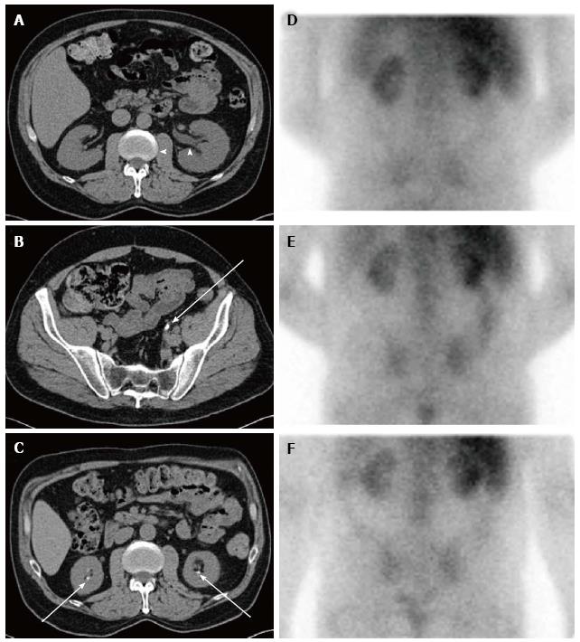
Figure 2 Sequential imaging studies of a not yet reported patient with chronic kidney disease from dietary hyperoxaluria.
Axial computed tomography (CT) images obtained two years before the hyperoxaluria diagnosis show (A) mild left hydronephrosis (arrowheads) caused by (B) a left distal ureteral calculus (arrow). Axial CT image obtained around the time of the hyperoxaluria diagnosis shows (C) bilateral nephrolithiasis (arrows). Nuclear medicine gallium-67 citrate scan images were also obtained around the time of diagnosis, including (D) 4-, (E) 24-, and (F) 48 h after administration. These show abnormal, persistent bilateral renal activity at all time points, indicative of interstitial nephritis. Gallium scanning has classically been used to distinguish acute interstitial nephritis from acute tubular necrosis and other causes of acute renal failure[216-218]. In this patient chronic interstitial nephritis associated with hyperoxaluria led to this positive scan. The patient’s diet for several years was based on nuts with estimated oxalate consumption ≥ 800 mg daily. During high oxalate intake, urine oxalate excretion was > 200 mg/24-h in several measurements obtained at serum creatinine levels > 3.5 mg/dL. After resumption of a diet low in oxalate and improvement of renal function to serum creatinine levels < 3.0 mg/dL, urine oxalate excretion decreased to normal levels.
- Citation: Glew RH, Sun Y, Horowitz BL, Konstantinov KN, Barry M, Fair JR, Massie L, Tzamaloukas AH. Nephropathy in dietary hyperoxaluria: A potentially preventable acute or chronic kidney disease. World J Nephrol 2014; 3(4): 122-142
- URL: https://www.wjgnet.com/2220-6124/full/v3/i4/122.htm
- DOI: https://dx.doi.org/10.5527/wjn.v3.i4.122


