Copyright
©The Author(s) 2016.
World J Virol. Aug 12, 2016; 5(3): 125-134
Published online Aug 12, 2016. doi: 10.5501/wjv.v5.i3.125
Published online Aug 12, 2016. doi: 10.5501/wjv.v5.i3.125
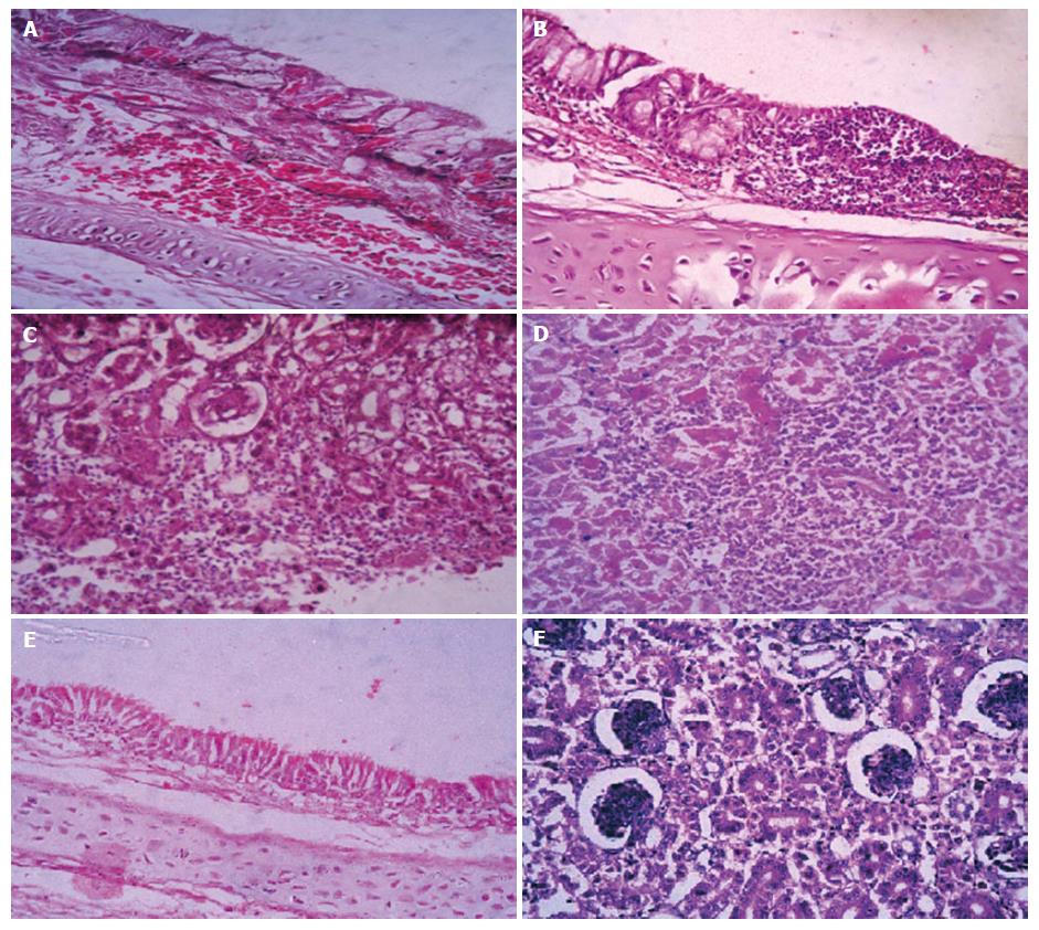
Figure 2 Histopathology illustration of the trachea and kidney from experimentally infected chickens.
A: Trachea (from Group 1) showed subepithelial hemorrhage accompanied with goblet cell activation, inflammatory cells and edema; B: Trachea (from Group 2) with focal aggregation of lymphocytic cells and epithelial desquamation, ulceration accompanied with goblet cell hypertrophy; C: Kidney (from Group 2) showed glomerular edema and glomerulonephritis with extravasation of blood vessels between renal tubules; D: Kidney (from Group 1) with severe necrosis of renal tubules and focal lymphocytic aggregation; E: Trachea of negative control group; F: Kidney of negative control group (H and E, × 20).
- Citation: Zanaty A, Arafa AS, Hagag N, El-Kady M. Genotyping and pathotyping of diversified strains of infectious bronchitis viruses circulating in Egypt. World J Virol 2016; 5(3): 125-134
- URL: https://www.wjgnet.com/2220-3249/full/v5/i3/125.htm
- DOI: https://dx.doi.org/10.5501/wjv.v5.i3.125


