Copyright
©2013 Baishideng Publishing Group Co.
World J Otorhinolaryngol. Nov 28, 2013; 3(4): 114-133
Published online Nov 28, 2013. doi: 10.5319/wjo.v3.i4.114
Published online Nov 28, 2013. doi: 10.5319/wjo.v3.i4.114
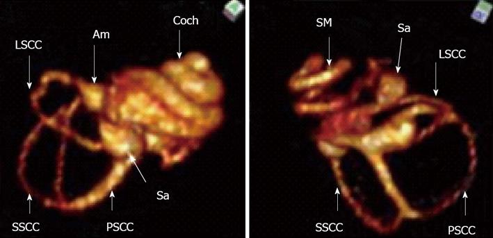
Figure 10 Magnetic resonance imaging of the inner ear of rat using SPION as contrast agent.
The upper figure shows T1 weighted imaging where all cochlear fluid spaces are visible. The lower figure shows T2 weighted image where SPION injected into the perilymphatic space will reduce the T2 signal and the perilymphatic space signal is extinguished. Only the fluid in scala media (endolymph) is visible. Am: Ampulla; Coch: Cochlea; Sa: Saccus; LSCC: Lateral semicircular canal; SSCC: Superior semicircular cananal; PSCC: Posterior semicicrcular canal; SM: Scala media.
- Citation: Pyykkö I, Zou J, Zhang Y, Zhang W, Feng H, Kinnunen P. Nanoparticle based inner ear therapy. World J Otorhinolaryngol 2013; 3(4): 114-133
- URL: https://www.wjgnet.com/2218-6247/full/v3/i4/114.htm
- DOI: https://dx.doi.org/10.5319/wjo.v3.i4.114


