Published online May 18, 2017. doi: 10.5312/wjo.v8.i5.379
Peer-review started: October 28, 2016
First decision: December 13, 2016
Revised: February 3, 2017
Accepted: March 12, 2017
Article in press: March 13, 2017
Published online: May 18, 2017
To describe an approach to anterior cruciate ligament (ACL) reconstruction using autologous hamstring by drilling via the anteromedial portal in the presence of an intramedullary (IM) femoral nail.
Once preoperative imagining has characterized the proposed location of the femoral tunnel preparations are made to remove all of the hardware (locking bolts and IM nail). A diagnostic arthroscopy is performed in the usual fashion addressing all intra-articular pathology. The ACL remnant and lateral wall soft tissues are removed from the intercondylar, to provide adequate visualization of the ACL footprint. Femoral tunnel placement is performed using a transportal ACL guide with desired offset and the knee flexed to 2.09 rad. The Beath pin is placed through the guide starting at the ACL’s anatomic footprint using arthroscopic visualization and/or fluoroscopic guidance. If resistance is met while placing the Beath pin, the arthroscopy should be discontinued and the obstructing hardware should be removed under fluoroscopic guidance. When the Beath pin is successfully placed through the lateral femur, it is overdrilled with a 4.5 mm Endobutton drill. If the Endobutton drill is obstructed, the obstructing hardware should be removed under fluoroscopic guidance. In this case, the obstruction is more likely during Endobutton drilling due to its larger diameter and increased rigidity compared to the Beath pin. The femoral tunnel is then drilled using a best approximation of the graft’s outer diameter. We recommend at least 7 mm diameter to minimize the risk of graft failure. Autologous hamstring grafts are generally between 6.8 and 8.6 mm in diameter. After reaming, the knee is flexed to 1.57 rad, the arthroscope placed through the anteromedial portal to confirm the femoral tunnel position, referencing the posterior wall and lateral cortex. For a quadrupled hamstring graft, the gracilis and semitendinosus tendons are then harvested in the standard fashion. The tendons are whip stitched, quadrupled and shaped to match the diameter of the prepared femoral tunnel. If the diameter of the patient’s autologous hamstring graft is insufficient to fill the prepared femoral tunnel, the autograft may be supplemented with an allograft. The remainder of the reconstruction is performed according to surgeon preference.
The presence of retained hardware presents a challenge for surgeons treating patients with knee instability. In cruciate ligament reconstruction, distal femoral and proximal tibial implants hardware may confound tunnel placement, making removal of hardware necessary, unless techniques are adopted to allow for anatomic placement of the graft.
This report demonstrates how the femoral tunnel can be created using the anteromedial portal instead of a transtibial approach for reconstruction of the ACL.
Core tip: The presence of retained hardware presents a challenge for surgeons treating patients with knee instability. In anterior cruciate ligament (ACL) reconstruction, intramedullary (IM) nails may confound tunnel placement, making removal of hardware necessary, unless techniques are adopted to allow for anatomic placement of the graft. We strongly recommend delaying the ACL graft harvest until creation of the femoral tunnel has been successful in these settings. Although unlikely when using anteromedial portal drilling, if the IM rod needs to be removed for anatomic graft placement but cannot be removed, the ACL reconstruction may have to be delayed until this issue is addressed.
- Citation: Lacey M, Lamplot J, Walley KC, DeAngelis JP, Ramappa AJ. Technical note: Anterior cruciate ligament reconstruction in the presence of an intramedullary femoral nail using anteromedial drilling. World J Orthop 2017; 8(5): 379-384
- URL: https://www.wjgnet.com/2218-5836/full/v8/i5/379.htm
- DOI: https://dx.doi.org/10.5312/wjo.v8.i5.379
Anterior cruciate ligament (ACL) reconstruction offers patients with knee instability an excellent result following an isolated ACL rupture. However, because this injury often occurs in conjunction with lower extremity trauma, ACL reconstruction may follow surgical fixation of femur and/or tibia fractures[1-5]. When the hardware is located in the distal femur or proximal tibia, it may obstruct the normal placement of the tibial or femoral tunnels. Preoperative planning and intraoperative fluoroscopy can facilitate anatomic placement of the femoral tunnel using the anteromedial portal (AMP) rather than a transtibial (TT) approach in order to avoid removal of retained hardware. It has been shown that the use of AMP may be superior to the TT drilling technique in the setting of acute ACL reonstruction based on physical examination and patient reported outcomes; however these reported improvements have neither reached a minimally clinically important difference nor have been reported in the setting of a femoral fixation hardware[6]. In this technical note, we describe an approach to ACL reconstruction using autologous hamstring by drilling via the AMP in the presence of an intramedullary (IM) femoral nail.
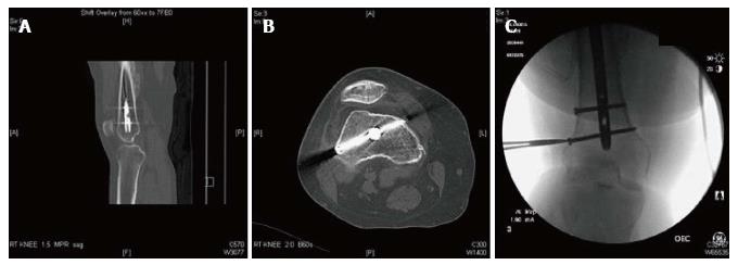
Preoperative planning: Preoperative imaging including a computed tomography (CT) scan of the distal femur is reviewed to assess the proposed location of the femoral tunnel (Figure 1A and B). Preparations are made to remove all of the hardware (locking bolts and IM nail) by requesting proper instrumentation, personnel and imaging support. While this process confirms that drilling via the AMP should avoid the IM nail, we recommend preparing the femoral tunnel before harvesting the hamstring tendons and preparing the graft after femoral drilling has been successfully completed in cases where the size of the femoral tunnel is a concern. Finally, since the femoral tunnel is drilled before harvesting autologous hamstring graft, a cadaveric graft should be available in case the diameter of the harvested hamstrings is insufficient to fill the femoral tunnel.
A diagnostic arthroscopy is performed in the usual fashion. All intra-articular pathology, including meniscal tears and loose bodies, is addressed. The ACL remnant and lateral wall soft tissues are removed from the intercondylar, to provide adequate visualization of the ACL footprint. Femoral tunnel placement is performed using a transportal ACL guide with desired offset (Arthrex, Naples, FL) and the knee flexed to 2.09 rad. The Beath pin is placed through the guide starting at the ACL’s anatomic footprint using arthroscopic visualization and/or fluoroscopic guidance. If resistance is met while placing the Beath pin, the arthroscopy should be discontinued and the obstructing hardware should be removed under fluoroscopic guidance. When the Beath pin is successfully placed through the lateral femur, it is overdrilled with a 4.5 mm Endobutton drill (Smith and Nephew, Andover, MA). If the Endobutton drill is obstructed, the obstructing hardware should be removed under fluoroscopic guidance (Figure 1C). In this case, the obstruction is more likely during Endobutton drilling due to its larger diameter and increased rigidity compared to the Beath pin. The femoral tunnel is then drilled using a best approximation of the graft’s outer diameter. We recommend at least 7 mm diameter to minimize the risk of graft failure[7]. Autologous hamstring grafts are generally between 6.8 and 8.6 mm in diameter[8]. After reaming, the knee is flexed to 1.57 rad, the arthroscope placed through the anteromedial portal to confirm the femoral tunnel position, referencing the posterior wall and lateral cortex.
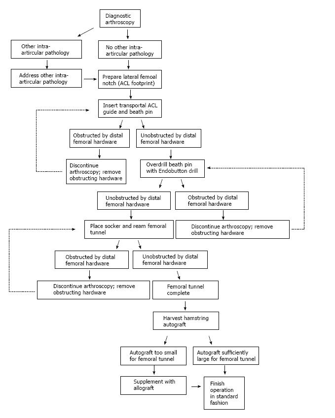
For a quadrupled hamstring graft, the gracilis and semitendinosus tendons are then harvested in the standard fashion. The tendons are whip stitched, quadrupled and shaped to match the diameter of the prepared femoral tunnel. If the diameter of the patient’s autologous hamstring graft is insufficient to fill the prepared femoral tunnel, the autograft may be supplemented with an allograft. The remainder of the reconstruction is performed according to surgeon preference (Figure 2).
We present a systematic approach to ACL reconstruction in the presence of distal femoral hardware using anteromedial portal femoral drilling followed by autologous hamstring harvest. Like several techniques of femoral tunneling, AMP drilling may provide improved rotation stability, decreased anterior translation and greater coverage of ACL’s anatomic footprint compared to TT techniques, but there is little evidence to support a clinical difference[6,9-12]. To this end, clinical outcomes of TT and AMP drilling techniques for ACL reconstruction were directly appraised in a 2016 systematic literature review, however all outcomes suggesting superior result of AMP drilling technique failed to surpass a minimal clinically important difference despite notable improvements based on the physical exam and scoring system results[6].
In a biomechanical setting, Steiner et al[13] argued that single-bundle ACL reconstructions may be improved if grafts are centered in their anatomical insertions by an independent drilling method vs grafts placed by a conventional TT drilling method. The proposed advantage of AMP femoral drilling is the creation of an independent tunnel, which may be oriented to avoid existing hardware. This benefit, depending on the location of the hardware as obstruction, may be unattainable. Ideally, this difficulty would be determined during preoperative planning, as outlined in (Table 1), using CT imaging.
| Obtain and review radiographic studies including computed tomography scan of distal femur to determine location of hardware which may interfere with femoral tunnel placement |
| Discuss feasibility and necessity of hardware removal, considering location of individual components and entire construct relative to planned femoral tunnel site, with primary surgeon or consulting trauma surgeon |
| Arrange for proper instrumentation, fluoroscopy and personnel for removal of hardware |
| Arrange for access to allograft in case hamstring autograft is insufficient in diameter |
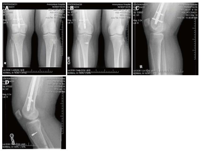
In this case, one distal locking screw was located approximately 2 cm superior to the intercondylar notch, adjacent to posterior femoral cortex and oriented from posterolateral to anteromedial (Figure 1). This screw had to be removed after an unsuccessful attempt at overdrilling the Beath pin (Figure 3). AMP drilling may allow the surgeon to minimize the amount of hardware removed. Because TT femoral drilling techniques result in a more vertically-oriented femoral tunnel that is closer to the midline in the coronal plane. Removal of multiple screws or the entire IM nail may have been necessary.
We strongly recommend delaying the hamstring harvest until creation of the femoral tunnel has been successful. Although unlikely when using AMP drilling, if the retained hardware needs to be removed but this process is unsuccessful, the ACL reconstruction may have to be delayed until this issue is addressed.
Anterior cruciate ligament (ACL) reconstruction offers patients with knee instability an excellent result following an isolated ACL rupture. However, because this injury often occurs in conjunction with lower extremity trauma, ACL reconstruction may follow surgical fixation of femur and/or tibia fractures.
When the hardware is located in the distal femur or proximal tibia, it may obstruct the normal placement of the tibial or femoral tunnels. Preoperative planning and intraoperative fluoroscopy can facilitate anatomic placement of the femoral tunnel using the anteromedial portal (AMP) rather than a transtibial (TT) approach in order to avoid removal of retained hardware.
It has been shown that the use of AMP was superior to the TT drilling technique in the setting of acute ACL reconstruction based on physical examination and patient reported outcomes, however this has not been reported in the setting of a femoral nail.
The authors strongly recommend delaying the hamstring harvest until creation of the femoral tunnel has been successful. Although unlikely when using AMP drilling, if the retained hardware needs to be removed but this process is unsuccessful, the ACL reconstruction may have to be delayed until this issue is addressed.
This is a short communication with a clear and useful message to other clinicians regarding the best approach to repair ACL injury whilst allowing correct positioning of other implant materials to repair local bone areas.
Manuscript source: Invited manuscript
Specialty type: Orthopedics
Country of origin: United States
Peer-review report classification
Grade A (Excellent): A
Grade B (Very good): B, B, B
Grade C (Good): 0
Grade D (Fair): 0
Grade E (Poor): 0
P- Reviewer: Cartmell S, Fenichel I, Palmieri-Smith RM, Peng BG S- Editor: Qi Y L- Editor: A E- Editor: Lu YJ
| 1. | Emami Meybodi MK, Ladani MJ, Emami Meybodi T, Rahimnia A, Dorostegan A, Abrisham J, Yarbeygi H. Concomitant ligamentous and meniscal knee injuries in femoral shaft fracture. J Orthop Traumatol. 2014;15:35-39. [PubMed] [DOI] [Cited in This Article: ] [Cited by in Crossref: 8] [Cited by in F6Publishing: 3] [Article Influence: 0.3] [Reference Citation Analysis (0)] |
| 2. | LaFrance RM, Gorczyca JT, Maloney MD. Anterior cruciate ligament reconstruction failure after tibial shaft malunion. Orthopedics. 2012;35:e267-e271. [PubMed] [DOI] [Cited in This Article: ] [Cited by in Crossref: 2] [Cited by in F6Publishing: 3] [Article Influence: 0.3] [Reference Citation Analysis (0)] |
| 3. | Parvari R, Ziv E, Lantner F, Heller D, Schechter I. Somatic diversification of chicken immunoglobulin light chains by point mutations. Proc Natl Acad Sci USA. 1990;87:3072-3076. [PubMed] [DOI] [Cited in This Article: ] [Cited by in Crossref: 11] [Cited by in F6Publishing: 12] [Article Influence: 0.4] [Reference Citation Analysis (0)] |
| 4. | Sharma G, Naik VA, Pankaj A. Displaced osteochondral fracture of the lateral femoral condyle associated with an acute anterior cruciate ligament avulsion fracture: a corollary of “the lateral femoral notch sign”. Knee Surg Sports Traumatol Arthrosc. 2012;20:1599-1602. [PubMed] [DOI] [Cited in This Article: ] [Cited by in Crossref: 10] [Cited by in F6Publishing: 10] [Article Influence: 0.8] [Reference Citation Analysis (0)] |
| 5. | Spiro AS, Regier M, Novo de Oliveira A, Vettorazzi E, Hoffmann M, Petersen JP, Henes FO, Demuth T, Rueger JM, Lehmann W. The degree of articular depression as a predictor of soft-tissue injuries in tibial plateau fracture. Knee Surg Sports Traumatol Arthrosc. 2013;21:564-570. [PubMed] [DOI] [Cited in This Article: ] [Cited by in Crossref: 38] [Cited by in F6Publishing: 38] [Article Influence: 3.5] [Reference Citation Analysis (0)] |
| 6. | Liu A, Sun M, Ma C, Chen Y, Xue X, Guo P, Shi Z, Yan S. Clinical outcomes of transtibial versus anteromedial drilling techniques to prepare the femoral tunnel during anterior cruciate ligament reconstruction. Knee Surg Sports Traumatol Arthrosc. 2015; Epub ahead of print. [PubMed] [DOI] [Cited in This Article: ] [Cited by in Crossref: 38] [Cited by in F6Publishing: 38] [Article Influence: 5.4] [Reference Citation Analysis (0)] |
| 7. | Magnussen RA, Lawrence JT, West RL, Toth AP, Taylor DC, Garrett WE. Graft size and patient age are predictors of early revision after anterior cruciate ligament reconstruction with hamstring autograft. Arthroscopy. 2012;28:526-531. [PubMed] [DOI] [Cited in This Article: ] [Cited by in Crossref: 420] [Cited by in F6Publishing: 424] [Article Influence: 35.3] [Reference Citation Analysis (0)] |
| 8. | Treme G, Diduch DR, Billante MJ, Miller MD, Hart JM. Hamstring graft size prediction: a prospective clinical evaluation. Am J Sports Med. 2008;36:2204-2209. [PubMed] [DOI] [Cited in This Article: ] [Cited by in Crossref: 110] [Cited by in F6Publishing: 116] [Article Influence: 7.3] [Reference Citation Analysis (0)] |
| 9. | Franceschi F, Papalia R, Rizzello G, Del Buono A, Maffulli N, Denaro V. Anteromedial portal versus transtibial drilling techniques in anterior cruciate ligament reconstruction: any clinical relevance? A retrospective comparative study. Arthroscopy. 2013;29:1330-1337. [PubMed] [DOI] [Cited in This Article: ] [Cited by in Crossref: 71] [Cited by in F6Publishing: 72] [Article Influence: 6.5] [Reference Citation Analysis (0)] |
| 10. | Robert HE, Bouguennec N, Vogeli D, Berton E, Bowen M. Coverage of the anterior cruciate ligament femoral footprint using 3 different approaches in single-bundle reconstruction: a cadaveric study analyzed by 3-dimensional computed tomography. Am J Sports Med. 2013;41:2375-2383. [PubMed] [DOI] [Cited in This Article: ] [Cited by in Crossref: 33] [Cited by in F6Publishing: 35] [Article Influence: 3.2] [Reference Citation Analysis (0)] |
| 11. | Wang H, Fleischli JE, Zheng NN. Transtibial versus anteromedial portal technique in single-bundle anterior cruciate ligament reconstruction: outcomes of knee joint kinematics during walking. Am J Sports Med. 2013;41:1847-1856. [PubMed] [DOI] [Cited in This Article: ] [Cited by in Crossref: 70] [Cited by in F6Publishing: 75] [Article Influence: 6.8] [Reference Citation Analysis (0)] |
| 12. | Azboy I, Demirtaş A, Gem M, Kıran S, Alemdar C, Bulut M. A comparison of the anteromedial and transtibial drilling technique in ACL reconstruction after a short-term follow-up. Arch Orthop Trauma Surg. 2014;134:963-969. [PubMed] [DOI] [Cited in This Article: ] [Cited by in Crossref: 27] [Cited by in F6Publishing: 27] [Article Influence: 2.7] [Reference Citation Analysis (0)] |
| 13. | Steiner ME, Battaglia TC, Heming JF, Rand JD, Festa A, Baria M. Independent drilling outperforms conventional transtibial drilling in anterior cruciate ligament reconstruction. Am J Sports Med. 2009;37:1912-1919. [PubMed] [DOI] [Cited in This Article: ] [Cited by in Crossref: 151] [Cited by in F6Publishing: 153] [Article Influence: 10.2] [Reference Citation Analysis (0)] |


