Copyright
©2013 Baishideng Publishing Group Co.
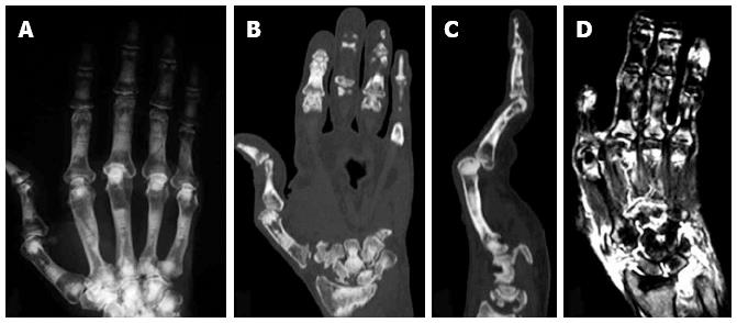
Figure 3 Plain X ray (A), computed tomography scan (B and C) and magnetic resonance imaging (D) of the right hand of an autosomal dominant II patient with rheumatoid arthritis.
Erosion of the carpal bones (B) and severe synovitis, as determined by the high intensity areas by T2-weighted magnetic resonance imaging images (D), were observed[14].
- Citation: Tanaka S. Regulation of bone destruction in rheumatoid arthritis through RANKL-RANK pathways. World J Orthop 2013; 4(1): 1-6
- URL: https://www.wjgnet.com/2218-5836/full/v4/i1/1.htm
- DOI: https://dx.doi.org/10.5312/wjo.v4.i1.1


