Copyright
©The Author(s) 2015.
World J Radiol. May 28, 2015; 7(5): 104-109
Published online May 28, 2015. doi: 10.4329/wjr.v7.i5.104
Published online May 28, 2015. doi: 10.4329/wjr.v7.i5.104
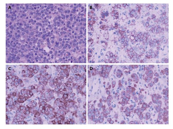
Figure 5 A biopsy of the prostate mass.
Microscopic findings (A) showed infiltrating nests of small cells in fibrotic stroma. Tumor cells had small hyperchromatic nuclei and scanty cytoplasms (H and E, × 200). Immunohistochemical stain showed strong positivity of tumor cells for CD56(++) (B), CgA(++) (C), Syn(++) (D).
- Citation: He HQ, Fan SF, Xu Q, Chen ZJ, Li Z. Diagnosis of prostatic neuroendocrine carcinoma: Two cases report and literature review. World J Radiol 2015; 7(5): 104-109
- URL: https://www.wjgnet.com/1949-8470/full/v7/i5/104.htm
- DOI: https://dx.doi.org/10.4329/wjr.v7.i5.104


