Copyright
©2012 Baishideng Publishing Group Co.
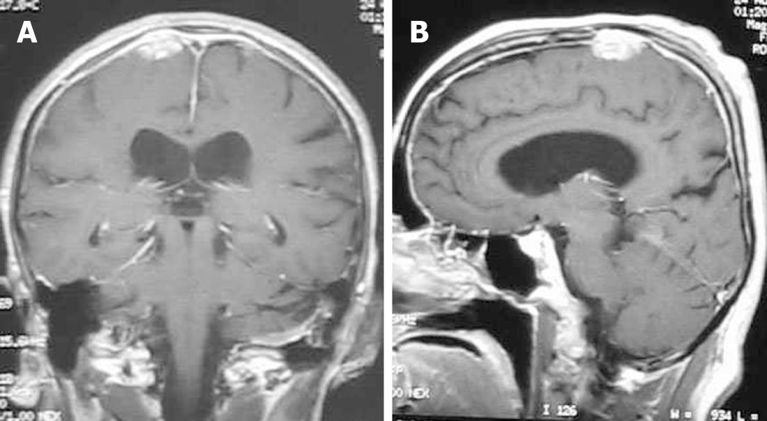
Figure 8 Neurosarcoidosis.
Coronal (A) and sagittal (B) T1-weighted magnetic resonance post-contrast images show an enhancing dural-based lesion. Also, note the enhancement of the dura along the convexity.
- Citation: Chourmouzi D, Potsi S, Moumtzouoglou A, Papadopoulou E, Drevelegas K, Zaraboukas T, Drevelegas A. Dural lesions mimicking meningiomas: A pictorial essay. World J Radiol 2012; 4(3): 75-82
- URL: https://www.wjgnet.com/1949-8470/full/v4/i3/75.htm
- DOI: https://dx.doi.org/10.4329/wjr.v4.i3.75


