Copyright
©The Author(s) 2018.
World J Radiol. Nov 28, 2018; 10(11): 172-183
Published online Nov 28, 2018. doi: 10.4329/wjr.v10.i11.172
Published online Nov 28, 2018. doi: 10.4329/wjr.v10.i11.172
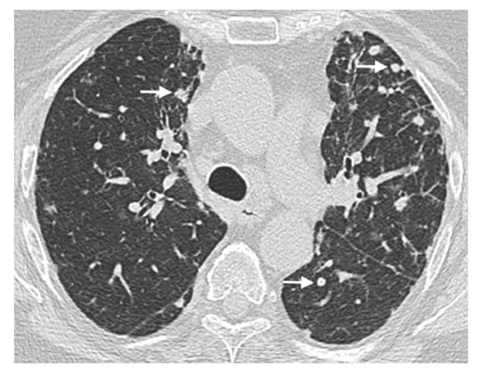
Figure 2 Nodules.
A 64-year-old female patient with common variable immunodeficiency disorder. High-resolution computed tomography shows multiple, small nodules with peribronchial and perifissural location in the upper and lower lobes of both lungs (arrows). A diagnosis of granulomatous and lymphocytic interstitial lung disease was subsequently made.
- Citation: Cereser L, De Carli M, d’Angelo P, Zanelli E, Zuiani C, Girometti R. High-resolution computed tomography findings in humoral primary immunodeficiencies and correlation with pulmonary function tests. World J Radiol 2018; 10(11): 172-183
- URL: https://www.wjgnet.com/1949-8470/full/v10/i11/172.htm
- DOI: https://dx.doi.org/10.4329/wjr.v10.i11.172


