Copyright
©The Author(s) 2017.
World J Hepatol. Nov 8, 2017; 9(31): 1205-1209
Published online Nov 8, 2017. doi: 10.4254/wjh.v9.i31.1205
Published online Nov 8, 2017. doi: 10.4254/wjh.v9.i31.1205
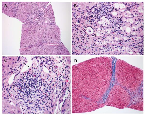
Figure 2 Resolving hepatitis.
A: The liver shows a somewhat nodular architecture with increased portal inflammatory cells, HE, × 100; B: Ballooned hepatocytes (arrow) and numerous pigmented Kupffer cells (asterix) are present in portal tracts consistent with injury, HE, × 400; C: Prominent eosinophils (arrow) suggests a drug reaction of the hypersensitivity type, HE, × 400; D: Bridging fibrosis (arrow). Thin fibrous bridges connect portal tracts, Trichrome, × 100.
- Citation: Dalal KK, Holdbrook T, Peikin SR. Ayurvedic drug induced liver injury. World J Hepatol 2017; 9(31): 1205-1209
- URL: https://www.wjgnet.com/1948-5182/full/v9/i31/1205.htm
- DOI: https://dx.doi.org/10.4254/wjh.v9.i31.1205


