Copyright
©2011 Baishideng Publishing Group Co.
World J Hepatol. Sep 27, 2011; 3(9): 256-261
Published online Sep 27, 2011. doi: 10.4254/wjh.v3.i9.256
Published online Sep 27, 2011. doi: 10.4254/wjh.v3.i9.256
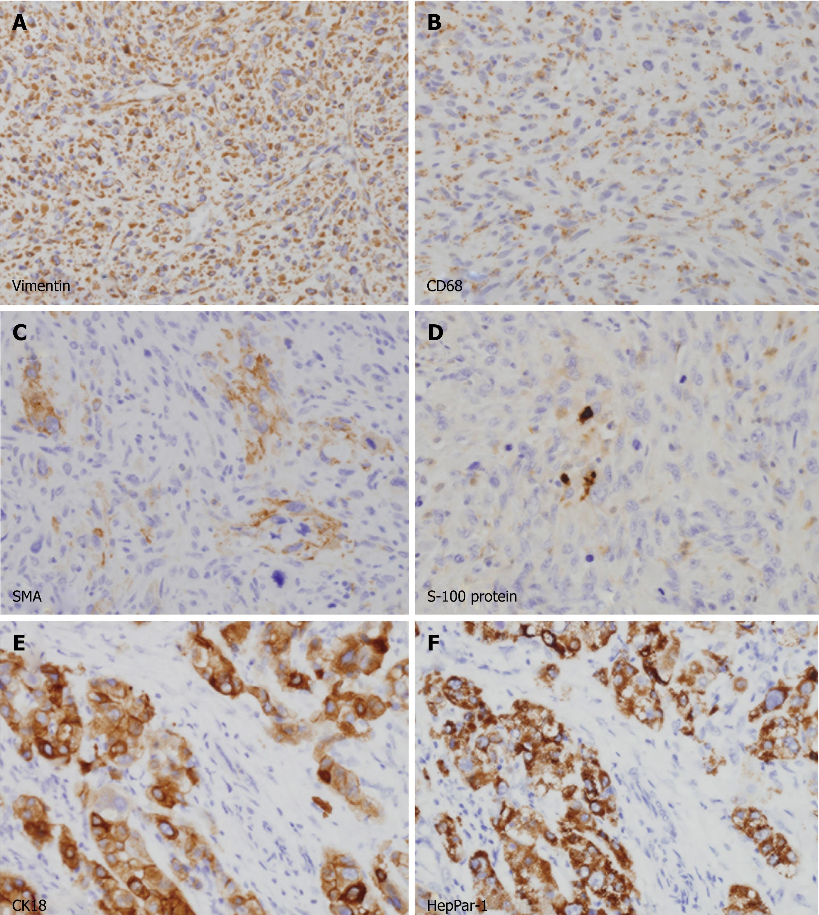
Figure 3 Immunohistochemical analyses of the tumors.
A-D: Spindle cells in the large mass were diffusely immunopositive for vimentin (A) and CD68 (B) in their cytoplasm. A few cells (less than 5%) were also positive for α-smooth muscle actin (C) and S-100 protein (D). All cells were negative for cytokeratins, hepatocyte paraffin-1 antigen (HepPar-1), desmin and myogenic differentiation-1, excluding cells of epithelial and myogenic differentiation (not shown); E, F: Polygonal cells in small mass were strongly immunopositive for cytokeratin 18 (E) and HepPar-1 (F), consistent with a typical hepatocellular carcinoma.
- Citation: Hwang HS, Ha ND, Jeong YK, Suh JH, Choi HJ, Kim YM, Cha HJ. Simultaneous occurrence of malignant fibrous histiocytoma and hepatocellular carcinoma in cirrhotic liver: A case report. World J Hepatol 2011; 3(9): 256-261
- URL: https://www.wjgnet.com/1948-5182/full/v3/i9/256.htm
- DOI: https://dx.doi.org/10.4254/wjh.v3.i9.256


