Copyright
©The Author(s) 2021.
World J Hepatol. Mar 27, 2021; 13(3): 315-327
Published online Mar 27, 2021. doi: 10.4254/wjh.v13.i3.315
Published online Mar 27, 2021. doi: 10.4254/wjh.v13.i3.315
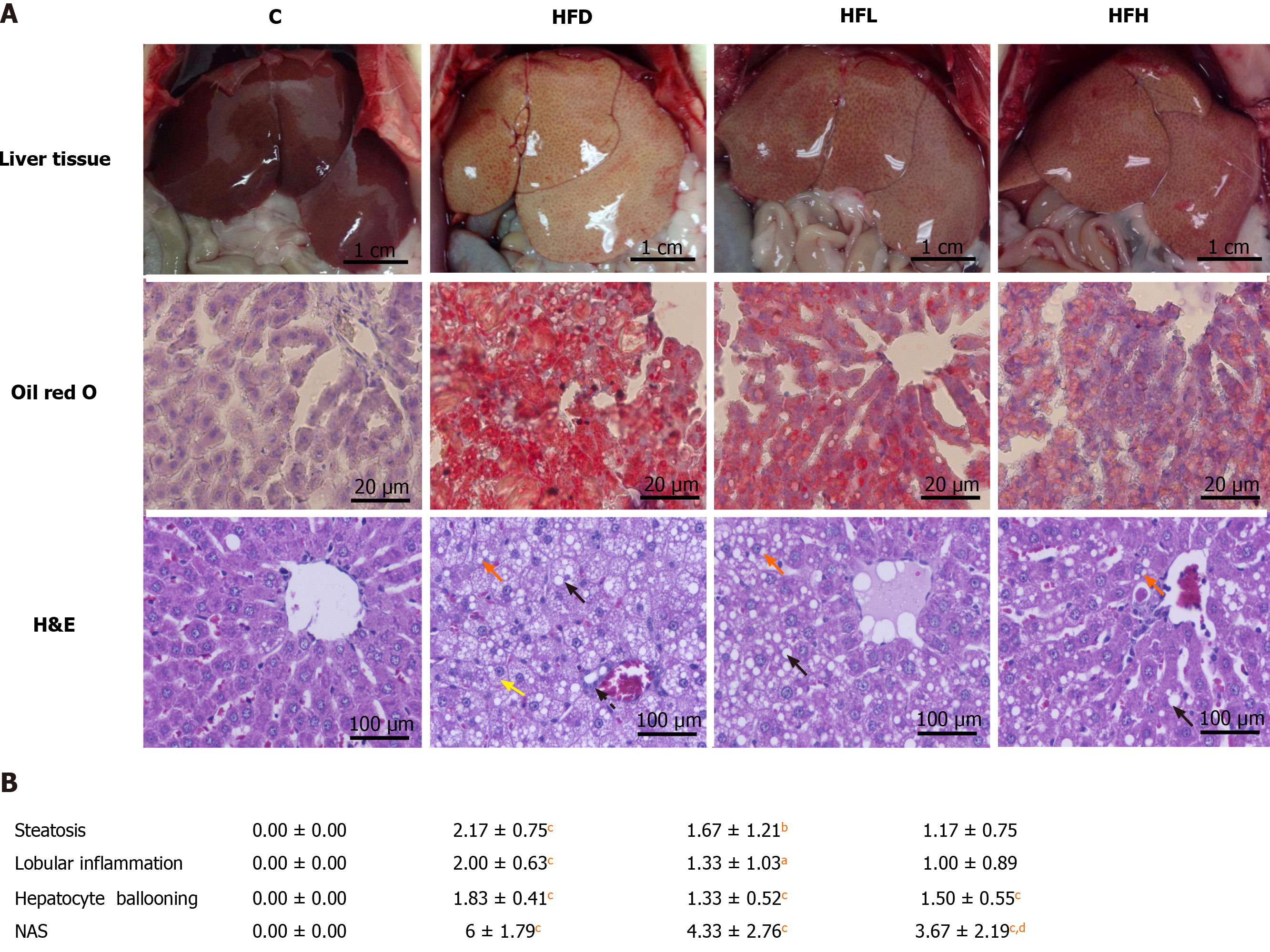
Figure 1 Effect of papaya on non-alcoholic fatty liver disease.
A: Macroscopic and microscopic appearance in rat hepatocytes. Macrovesicular steatosis (black arrow) are large lipid droplets that are present in the hepatocytes. Microvesicular steatosis (red arrow) are small lipid droplets that are present in the hepatocytes. Hepatocyte ballooning is recognised as cell swelling and enlargement within the cytoplasm (yellow arrow). Lobular inflammation in non-alcoholic steatohepatitis foci (dotted line arrow) are scattered in the hepatic lobule; B: Comparative analysis of non-alcoholic fatty liver disease activity score for all treatment groups. Data are expressed as mean ± SE of the mean (n = 6-7). aP < 0.05, bP < 0.01, cP < 0.001 vs C, and dP < 0.05 vs high fat diet group. C: Control; H&E: Hematoxylin and eosin; HFD: High fat diet; HFH: High fat diet treated with 1 mL of papaya juice/100 g body weight; HFL: High fat diet treated with 0.5 mL of papaya juice/100 g body weight; NAS: Non-alcoholic fatty liver disease activity score.
- Citation: Deenin W, Malakul W, Boonsong T, Phoungpetchara I, Tunsophon S. Papaya improves non-alcoholic fatty liver disease in obese rats by attenuating oxidative stress, inflammation and lipogenic gene expression. World J Hepatol 2021; 13(3): 315-327
- URL: https://www.wjgnet.com/1948-5182/full/v13/i3/315.htm
- DOI: https://dx.doi.org/10.4254/wjh.v13.i3.315


