Copyright
©The Author(s) 2019.
World J Stem Cells. Feb 26, 2019; 11(2): 84-99
Published online Feb 26, 2019. doi: 10.4252/wjsc.v11.i2.84
Published online Feb 26, 2019. doi: 10.4252/wjsc.v11.i2.84
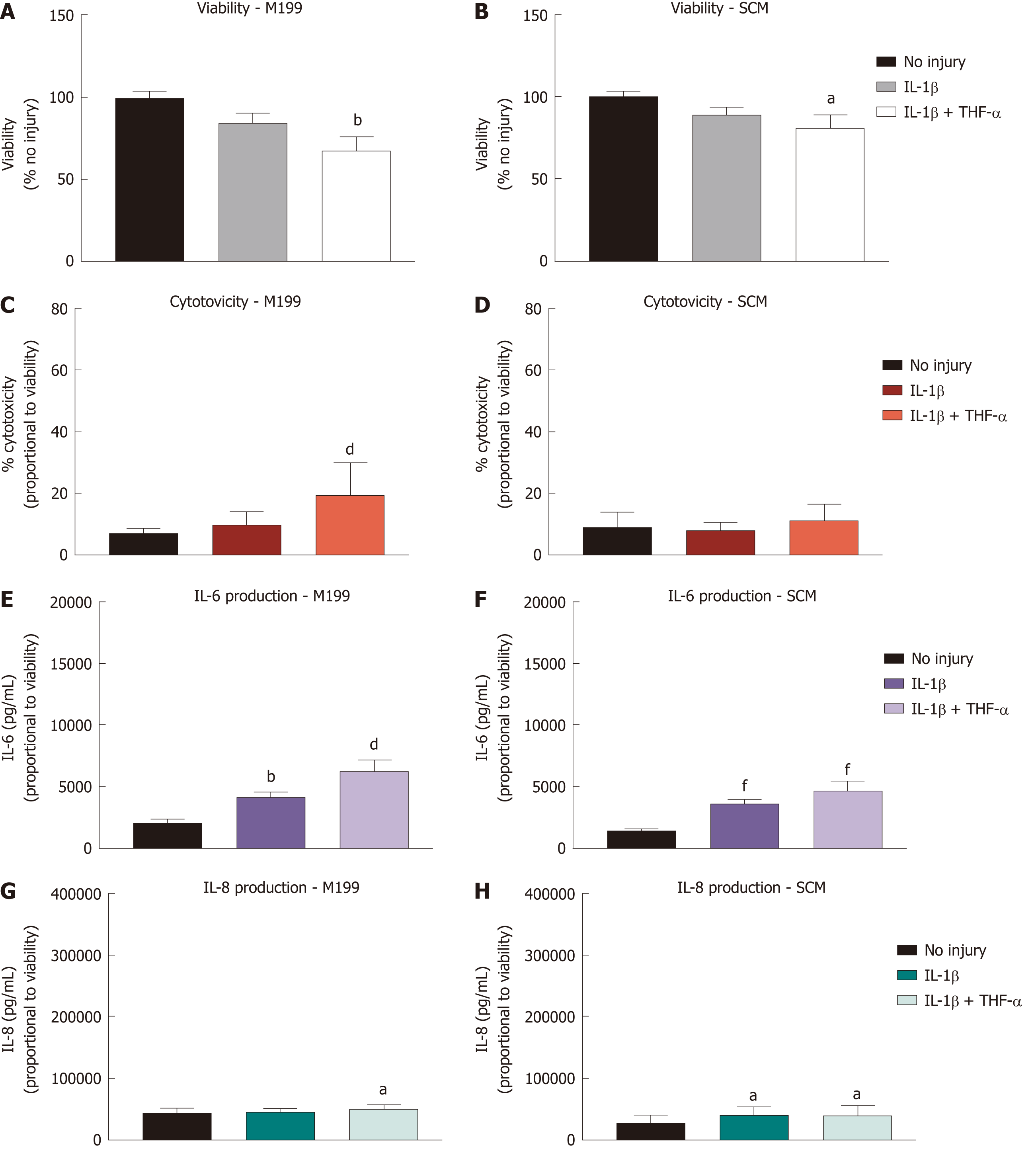
Figure 4 Effect of treatment with pro-inflammatory cytokines on corneal-stroma derived stem cells.
Treatment of corneal-stroma derived stem cells cultured in stem cell medium or M199 with 1 ng/mL IL-1β with/without 10 ng/mL tumour necrosis factor-α was performed for 72 h. A: PrestoBlue viability assay after 72 h stimulation. Data represented relative to reading for no injury control; B: Lactate dehydrogenase cytotoxicity assay performed on cell supernatants after 72 h treatment. Data displayed as percentage cytotoxicity and relative to cell viability; C: Concentration of IL-6 in the supernatant 72 h after injury. Data displayed relative to cell viability; D: Concentration of IL-8 in the supernatant 72 h after injury. Data displayed relative to cell viability. Data for all graphs shown as mean ± SEM of five independent experiments with three to six replicates each. Statistical significance compared to no injury controls analysed by one-way ANOVA represented by aP ≤ 0.05, bP ≤ 0.01, dP ≤ 0.001, fP ≤ 0.0001. IL: Interleukin; TNF: Tumour necrosis factor; SCM: Stem cell medium.
- Citation: Orozco Morales ML, Marsit NM, McIntosh OD, Hopkinson A, Sidney LE. Anti-inflammatory potential of human corneal stroma-derived stem cells determined by a novel in vitro corneal epithelial injury model. World J Stem Cells 2019; 11(2): 84-99
- URL: https://www.wjgnet.com/1948-0210/full/v11/i2/84.htm
- DOI: https://dx.doi.org/10.4252/wjsc.v11.i2.84


