Published online Apr 15, 2003. doi: 10.3748/wjg.v9.i4.710
Revised: October 10, 2002
Accepted: October 17, 2002
Published online: April 15, 2003
AIM: To investigate the role of glycylproline dipeptidyl aminopeptidase (GPDA) isoenzyme in the diagnosis of primary hepatocellular carcinoma (PHC), especially in patients with negative alpha-fetoprotein (AFP).
METHODS: A stage gradient polyacrylamide gel discontinuous electrophoresis system was developed to separate serum GPDA isoenzymes, which were determined in 102 patients with PHC, 45 cases with liver cirrhosis, 24 cases with chronic hepatitis, 35 cases with benign liver space-occupying lesions, 20 cases with metastatic liver cancer and 50 cases with extra-hepatic cancer, as well as 80 healthy subjects. The relationships between GPDA isoenzymes and AFP, the sizes of tumors, as well as alanine aminotransferase (ALT) were also analyzed.
RESULTS: Serum GPDA was separated into two isoenzymes, GPDA-S and GPDA-F. The former was positive in all subjects, while the latter was found mainly in majority of PHC (85.3%) and a few cases with liver cirrhosis (11.1%), chronic hepatitis (33.3%), metastatic liver cancer (15.0%) and non-hepatic cancer (16.0%). GPDA-F was negative in all healthy subjects and patients with benign liver space-occupying lesions, including abscess, cysts and angioma. There was no correlation between GPDA-F and AFP concentration or tumor size. GPDA-F was consistently positive and not correlated with ALT in PHC, but GPDA-F often converted to negative as decline of ALT in benign liver diseases. The electrophoretic migration of GPDA-F became sluggish after the treatment of neuraminidase.
CONCLUSION: GPDA-F is a new useful serum marker for PHC. Measurement of serum GPDA-F is helpful in diagnosis of PHC, especially in patients with negative AFP. GPDA-F is one kind of glycoproteins rich in sialic acid.
- Citation: Ni RZ, Huang JF, Xiao MB, Li M, Meng XY. Glycylproline dipeptidyl aminopeptidase isoenzyme in diagnosis of primary hepatocellular carcinoma. World J Gastroenterol 2003; 9(4): 710-713
- URL: https://www.wjgnet.com/1007-9327/full/v9/i4/710.htm
- DOI: https://dx.doi.org/10.3748/wjg.v9.i4.710
Glycylproline dipeptidyl aminopeptidase (GPDA) was discovered by Hapsu-Hava and Glenner in 1966[1]. It is widely distributed in human and animal tissues and catalyzes the hydrolysis of N-terminal glycylproline from glycylprolyl-β-naphthylamide and glycylproline-β-nitroanilide. It was reported that serum GPDA activity increased in the patients with hepatobiliary diseases and in experimental hepatic cancer[2,3]. Further studies demonstrated that GPDA activity was significantly higher in human hepatic cancer and embryonal tissues than in healthy adult liver tissues[4]. However, serum GPDA cannot be used as a marker for PHC due to its low sensitivity and specificity in diagnosis.
In order to improve the diagnostic value of GPDA for PHC, a stage gradient polyacrylamide gel discontinuous electrophoresis system was established to separate serum GPDA into two isoenzymes, and clinical significances of serum GPDA isoenzymes in diagnosis of PHC were studied.
The resources of all patients in this study were from Affiliated Hospital of Nantong Medical College. The diagnosis of PHC met the diagnostic criterion of Liver Cancer Branch Committee of Chinese Anti-cancer Association. The diagnosis in 35 patients was confirmed pathologically. The origins of 20 metastatic liver cancers were gastrointestinal tract cancer (16 cases), lung cancer (2 cases) and breast cancer (2 cases). The origins in 50 cases with extra-hepatic cancer were gastrointestinal tract (22 cases), pancreas (5 cases), bile duct (6 cases ), ovary (8 cases), bladder (4 cases), lung (4 cases) and kidney (1 case). Thirty-five benign liver space-occupying lesions were composed of 8 liver abscess, 2 liver cysts and 25 liver angioma. The diagnoses of 45 cases with hepatic cirrhosis and 24 chronic hepatitis were based on clinical manifestations and liver biopsy. The control samples came from healthy blood donor.
Separation of GPDA isoenzymes was performed on stage gradient polyacrylamide gel with a vertical slab electrophoresis apparatus. The gel consists of one layer of stack gel and two layers of separating gels. The following solutions were used for the preparation of gels: (1) Solution A: 30% acrylamide containing 0.8% bisacrylamide; (2) Solution B: 4.95% Tris base containing 0.05% TEMED, pH6.9; (3) Solution C: 0.14% ammonium persulfate. A gel of dimensions of 12.0 cm × 10.0 cm × 0.15 cm was prepared and loaded with 20 µl of serum. The gel run at a constant voltage of 60 V for the first three hours and then at the voltage of 100 V for another 16 hours. After the electrophoresis, the gel was incubated at 37 °C for 30 min with a piece of acetate cellulose membrane of the gel size pre-soaked with a substrate solution, which consists of 15 mg of glycylproline-β-nitroanilide, 4.8 ml of 100 mmol·L-1 Tris-glycylglycine (pH8.4), 0.1 ml of 100 ml·L-1 sodium nitrite and 0.1 ml of 50 g·L-1 N-(naphthyl)-ethylenediamine dihydrochloride. Finally red GPDA isoenzyme bands were visualized soon by immersing the acetate cellulose membrane into a solution composed of 100 ml·L-1 trichloracetic acid and 250 ml·L-1 glycerol.
ALT was measured with the method recommended by International Federation of Clinical Chemistry[5] and AFP was determined with routine radioimmunoassay.
Serum was incubated with 10 u/mL neuraminidase at 37 °C for 15 hr before electrophoresis.
Chi-square test and student t tests were used. P < 0.05 was considered as statistically significant.
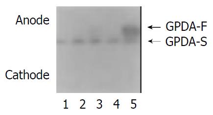
Serum GPDA was separated into two isoenzyme bands with the electrophoresis system (Figure 1). The band in the anode was called as GPDA-F (fast band) and the band in cathode as GPDA-S (slow band). GPDA-S was positive in all subjects, including healthy persons and patients, while positive rates of GPDA-F varied in different diseases. GPDA-F was found in most patients with PHC and a few patients with benign liver diseases and extra-hepatic cancer, but no GPDA-F was found in all healthy persons and the patients with benign liver space-occupying lesions (Table 1). In patients with PHC, positive GPDA-F was considered as true positive and negative GPDA-F as false negative, while in patients with other diseases positive GPDA-F was considered as false positive and negative GPDA-F as true negative. Therefore, the sensitivity, specificity and accuracy of GPDA-F in diagnosis of PHC were 85.3%, 86.2% and 85.9% respectively (Table 1).
| GPDA-F (+) | GPDA-F (-) | |||
| n | % | n | % | |
| Healthy subjects | 0 | 0 | 80 | 100.0 |
| PHC | 87a | 85.3 | 15 | 14.7 |
| Liver cirrhosis | 5 | 11.1 | 40 | 88.9 |
| Chronic hepatitis | 8 | 33.3 | 16 | 66.7 |
| Benign liver space- | 0 | 0 | 35 | 100.0 |
| occupying lesions | ||||
| Metastatic hepatic cancer | 3 | 15.0 | 17 | 85.0 |
| Extra-hepatic cancer | 8 | 16.0 | 42 | 84.0 |
There was no correlation between positive rates of GPDA-F and AFP concentration (Table 2). GPDA-F was positive in 75.0% PHC cases with negative AFP (< 50 ng/mL). If positive GPDA-F and AFP ≥ 50 ng/mL were set as diagnostic criterion of PHC, the sensitivity of AFP was 72.5% and that of AFP with GPDA-F would reach up to 91.2%. The diagnostic accuracy was also increased with the combination of AFP and GPDA-F (Table 3).
| AFP (ng/mL) | n | GPDA-F (+) |
| < 50 | 28 | 21 (75.0%) |
| -400 | 12 | 11 (91.6%) |
| > 400 | 62 | 55 (88.7%) |
| Sensitivity (%) | Specificity (%) | Accuracy (%) | |
| AFP alone | 72.5 | 95.2 | 85.0 |
| GPDA-F alone | 85.3 | 87.1 | 86.3 |
| AFP and GPDA-F | 91.2 | 84.7 | 88.5 |
One hundred and two cases with PHC were divided into two groups based on size of tumor, 23 cases were small liver cancer with tumor diameter ≤ 5 cm and 79 cases were advanced liver cancer with tumor diameter > 5 cm. Positive rate of GPDA-F was 78.3% (18/23) in small liver cancer, and 87.3% (69/79) in advanced liver cancer. However, there was no statistical difference between the two groups (P > 0.05).
In order to know whether GPDA-F is associated with inflammation of hepatocytes, GPDA-F and ALT were measured simultaneously. There was no significant difference of ALT activities between positive and negative GPDA-F groups in PHC, but ALT activities in positive GPDA-F group were significantly higher than that in negative group in liver cirrhosis and chronic hepatitis (Table 4), suggesting that GPDA-F was associated with elevated ALT in benign liver diseases, but not in PHC. It was also found that GPDA-F often converted to negative with the decrease of ALT in benign liver diseases but no in PHC (data not shown).
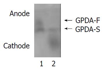
Serum was treated with neuraminidase before electrophoresis as mentioned in materials and methods. The migration rate of GPDA-F became slower than that of GPDA-S following treatment of neuraminidase, while GPDA-S was not obviously affected (Figure 2).
PHC is one of the most severe malignancies and ranks second mortality in malignant tumors in China[6,7]. The prognosis is poor due to its delayed diagnosis and low resectable rate. Thus early diagnosis is important to improve the prognosis. Tumor markers are very useful in diagnosis. AFP is the most widely used tumor marker for PHC with a positive rate from 60% to 70%[8-11]. AFP-L-3, one of the glycoforms of AFP, has higher specificity for PHC. However, it cannot increase the diagnostic sensitivity[12-16]. Many studies have focused on other tumor markers to improve the diagnostic sensitivity for PHC. It has been found that there are some tumor markers, which are complementary to AFP in diagnosis of PHC, such as des-gamma-carboxy prothrombin (PIVKA-II)[17-23], γ-glutamyltransferase isoenzyme[24-26], transforming growth factor-β1[27-29] and α-L-fucosidase[30-34].
In present study, we have investigated the value of GPDA isoenzymes in diagnosis of PHC. With a stage gradient polyacrylamide gel electrophoresis system, serum GPDA has been separated into two isoenzymes, GPDA-F and GPDA-S. GPDA-F was often detected in sera of patients with PHC, with a much higher positive rate than in other liver diseases, including liver cirrhosis, chronic hepatitis, metastatic liver cancer and extra-hepatic tumors; while GPDA-F was negative in healthy subjects and patients with benign liver space-occupying lesions. The sensitivity and specificity of GPDA-F in diagnosis of PHC were 85.3% and 86.2%, respectively. These results indicate that GPDA-F may be a valuable serum marker for PHC.
Serum GPDA-F in patients with benign liver diseases possibly originates from the inflammatory hepatocytes because in such cases positive GPDA-F was often accompanied by elevated ALT and changed to negative with the decline of ALT. On the contrary, GPDA-F in PHC is persistently positive and not correlated with ALT. Therefore, simultaneous determination and follow-up of ALT may help to rule out the false positive GPDA-F in benign liver diseases. It is known that some serum hepatoma markers are of predictive and early diagnostic value for PHC, such as AFP, des-gamma-carboxy prothrombin and gamma-glutamyltransferase isoenzyme II[21-23,26]. Whether persistent positive of GPDA-F in a few cases of liver cirrhosis is the precursor of hepatocarcinogenesis needs further investigation.
Although the diagnostic efficiency for PHC has been greatly improved in recent years with the use of many advanced image techniques, it is still difficult to make the diagnosis in some cases, especially for those at early stage or with negative AFP. Present study showed that GPDA-F and AFP were complementary for diagnosis of PHC. In these data, GPDA-F was positive in 75% PHC cases of negative AFP but negative in all benign liver space-occupying lesions. Therefore, GPDA-F is of special value for the differential diagnosis of liver space-occupying lesions of unknown nature. Our data also showed that there was no correlation between tumor size and GPDA-F. In 23 cases of small liver cancer, the positive rate of GPDA-F was 78.3%, suggesting that GPDA-F may be a good marker of PHC at early stage.
The exact mechanism by which GPDA-F appears in the sera of PHC cases is not clarified. It is possible that GPDA-F is produced by hepatoma tissues and then released into blood, since GPDA-F was only found in sera of those PHC cases in whose hepatoma tissues GPDA-F was positive (unpublished data). The electrophoretic migration of GPDA-F became slower than its isoenzyme GPDA-S after treatment with neuramidinase, indicating that GPDA-F is a kind of glycoprotein rich in sialic acids in its sugar chain. Therefore, the modification of the sugar chain after GPDA protein synthesis may be the main process of the production of GPDA-F. It has been known that several kinds of glycoproteins synthesized by hepatoma cells are different from those produced by hepatocytes. For example, AFP and alpha-1-antitrypsin have higher percentage of lens culinaris agglutinin-reactive species in hepatocellular carcinoma than in benign liver diseases. It is also clear that the fucosylation of the sugar chain at the innermost N-acetylglucosamine is the molecular basis of this variation[35,36] Whether there is the difference of fucosylation of GPDA isoenzymes between hepatocellular carcinoma and benign liver diseases needs to be studied further.
Edited by Ren SY
| 1. | Hopsu-Havu VK, Glenner GG. A new dipeptide naphthylamidase hydrolyzing glycyl-prolyl-beta-naphthylamide. Histochemie. 1966;7:197-201. [PubMed] [DOI] [Cited in This Article: ] [Cited by in Crossref: 316] [Cited by in F6Publishing: 318] [Article Influence: 5.5] [Reference Citation Analysis (0)] |
| 2. | Hutchinson DR, Halliwell RP, Lockhart JD, Parke DV. Glycylprolyl-p-nitroanilidase in hepatobiliary disease. Clin Chim Acta. 1981;109:83-89. [PubMed] [DOI] [Cited in This Article: ] [Cited by in Crossref: 9] [Cited by in F6Publishing: 9] [Article Influence: 0.2] [Reference Citation Analysis (0)] |
| 3. | Kojima J, Kanatani M, Nakamura N, Kashiwagi T, Tohjoh F, Akiyama M. Serum and liver glycylproline dipeptidyl aminopeptidase activity in rats with experimental hepatic cancer. Clin Chim Acta. 1980;107:105-110. [PubMed] [DOI] [Cited in This Article: ] [Cited by in Crossref: 8] [Cited by in F6Publishing: 8] [Article Influence: 0.2] [Reference Citation Analysis (0)] |
| 4. | Kojima J, Ueno Y, Kasugai H, Okuda S, Akedo H. Glycylproline dipeptidyl aminopeptidase and gamma-glutamyl transpeptidase in human hepatic cancer and embryonal tissues. Clin Chim Acta. 1987;167:285-291. [PubMed] [DOI] [Cited in This Article: ] [Cited by in Crossref: 13] [Cited by in F6Publishing: 13] [Article Influence: 0.4] [Reference Citation Analysis (0)] |
| 5. | Bergmeyer HU, Hørder M, Rej R. International Federation of Clinical Chemistry (IFCC) Scientific Committee, Analytical Section: approved recommendation (1985) on IFCC methods for the measurement of catalytic concentration of enzymes. Part 3. IFCC method for alanine aminotransferase (L-alanine: 2-oxoglutarate aminotransferase, EC 2.6.1.2). J Clin Chem Clin Biochem. 1986;24:481-495. [PubMed] [Cited in This Article: ] |
| 6. | Tang ZY. Hepatocellular carcinoma--cause, treatment and metastasis. World J Gastroenterol. 2001;7:445-454. [PubMed] [Cited in This Article: ] |
| 7. | Tang ZY. Hepatocellular carcinoma. J Gastroenterol Hepatol. 2000;15 Suppl:G1-G7. [PubMed] [DOI] [Cited in This Article: ] [Cited by in Crossref: 42] [Cited by in F6Publishing: 45] [Article Influence: 1.9] [Reference Citation Analysis (0)] |
| 8. | Rabe C, Pilz T, Klostermann C, Berna M, Schild HH, Sauerbruch T, Caselmann WH. Clinical characteristics and outcome of a cohort of 101 patients with hepatocellular carcinoma. World J Gastroenterol. 2001;7:208-215. [PubMed] [Cited in This Article: ] |
| 9. | Tangkijvanich P, Anukulkarnkusol N, Suwangool P, Lertmaharit S, Hanvivatvong O, Kullavanijaya P, Poovorawan Y. Clinical characteristics and prognosis of hepatocellular carcinoma: analysis based on serum alpha-fetoprotein levels. J Clin Gastroenterol. 2000;31:302-308. [PubMed] [DOI] [Cited in This Article: ] [Cited by in Crossref: 212] [Cited by in F6Publishing: 229] [Article Influence: 9.5] [Reference Citation Analysis (0)] |
| 10. | Yoshida S, Kurokohchi K, Arima K, Masaki T, Hosomi N, Funaki T, Murota M, Kita Y, Watanabe S, Kuriyama S. Clinical significance of lens culinaris agglutinin-reactive fraction of serum alpha-fetoprotein in patients with hepatocellular carcinoma. Int J Oncol. 2002;20:305-309. [PubMed] [Cited in This Article: ] |
| 11. | Qin LX, Tang ZY. The prognostic significance of clinical and pathological features in hepatocellular carcinoma. World J Gastroenterol. 2002;8:193-199. [PubMed] [Cited in This Article: ] |
| 12. | Tamura A, Oita T, Sakizono K, Nakajima T, Kasakura S. [Clinical usefulness of lectin-reactive fraction of alpha-fetoprotein in hepatocellular carcinoma]. Rinsho Byori. 1998;46:158-162. [PubMed] [Cited in This Article: ] |
| 13. | Zaninotto M, Ujka F, Lachin M, Bernardi D, Marafi C, Farinati F, Plebani M. Lectin-affinity electrophoresis for the detection of AFP microheterogeneities in patients with hepatocellular carcinoma. Anticancer Res. 1996;16:305-309. [PubMed] [Cited in This Article: ] |
| 14. | Kumada T, Nakano S, Takeda I, Kiriyama S, Sone Y, Hayashi K, Katoh H, Endoh T, Sassa T, Satomura S. Clinical utility of Lens culinaris agglutinin-reactive alpha-fetoprotein in small hepato-cellular carcinoma: special reference to imaging diagnosis. J Hepatol. 1999;30:125-130. [DOI] [Cited in This Article: ] [Cited by in Crossref: 106] [Cited by in F6Publishing: 112] [Article Influence: 4.5] [Reference Citation Analysis (0)] |
| 15. | Hayashi K, Kumada T, Nakano S, Takeda I, Sugiyama K, Kiriyama S, Sone Y, Miyata A, Shimizu H, Satomura S. Usefulness of measurement of Lens culinaris agglutinin-reactive fraction of alpha-fetoprotein as a marker of prognosis and recurrence of small hepatocellular carcinoma. Am J Gastroenterol. 1999;94:3028-3033. [PubMed] [Cited in This Article: ] |
| 16. | Li D, Mallory T, Satomura S. AFP-L3: a new generation of tumor marker for hepatocellular carcinoma. Clin Chim Acta. 2001;313:15-19. [PubMed] [DOI] [Cited in This Article: ] [Cited by in Crossref: 238] [Cited by in F6Publishing: 227] [Article Influence: 9.9] [Reference Citation Analysis (0)] |
| 17. | Sakon M, Monden M, Gotoh M, Kanai T, Umeshita K, Nakano Y, Mori T, Sakurai M, Wakasa K. Relationship between patho-logic prognostic factors and abnormal levels of des-gamma-carboxy prothrombin and alpha-fetoprotein in hepatocellular carcinoma. Am J Surg. 1992;163:251-256. [DOI] [Cited in This Article: ] [Cited by in Crossref: 28] [Cited by in F6Publishing: 30] [Article Influence: 0.9] [Reference Citation Analysis (0)] |
| 18. | Lamerz R, Runge M, Stieber P, Meissner E. Use of serum PIVKA-II (DCP) determination for differentiation between benign and malignant liver diseases. Anticancer Res. 1999;19:2489-2493. [PubMed] [Cited in This Article: ] |
| 19. | Tanaka Y, Kashiwagi T, Tsutsumi H, Nagasawa M, Toyama T, Ozaki S, Naito M, Ishibashi K, Azuma M. Sensitive measurement of serum abnormal prothrombin (PIVKA-II) as a marker of hepatocellular carcinoma. Hepatogastroenterology. 1999;46:2464-2468. [PubMed] [Cited in This Article: ] |
| 20. | Sassa T, Kumada T, Nakano S, Uematsu T. Clinical utility of simultaneous measurement of serum high-sensitivity des-gamma-carboxy prothrombin and lens culinaris agglutinit A-re-active alpha-fetoprotein in patients with small hepatocellular carcinoma. Eur J Gastroenterol Hepatol. 1999;11:1387-1392. [DOI] [Cited in This Article: ] [Cited by in Crossref: 51] [Cited by in F6Publishing: 55] [Article Influence: 2.2] [Reference Citation Analysis (0)] |
| 21. | Ishii M, Gama H, Chida N, Ueno Y, Shinzawa H, Takagi T, Toyota T, Takahashi T, Kasukawa R. Simultaneous measurements of serum alpha-fetoprotein and protein induced by vitamin K absence for detecting hepatocellular carcinoma. South Tohoku District Study Group. Am J Gastroenterol. 2000;95:1036-1040. [PubMed] [Cited in This Article: ] |
| 22. | Ikoma J, Kaito M, Ishihara T, Nakagawa N, Kamei A, Fujita N, Iwasa M, Tamaki S, Watanabe S, Adachi Y. Early diagnosis of hepatocellular carcinoma using a sensitive assay for serum des-gamma-carboxy prothrombin: a prospective study. Hepatogastroenterology. 2002;49:235-238. [PubMed] [Cited in This Article: ] |
| 23. | Fujiyama S, Tanaka M, Maeda S, Ashihara H, Hirata R, Tomita K. Tumor markers in early diagnosis, follow-up and management of patients with hepatocellular carcinoma. Oncology. 2002;62 Suppl 1:57-63. [PubMed] [DOI] [Cited in This Article: ] [Cited by in Crossref: 97] [Cited by in F6Publishing: 105] [Article Influence: 4.8] [Reference Citation Analysis (0)] |
| 24. | Okuyama K. Separation and identification of serum gamma-glutamyl transpeptidase isoenzymes by wheat germ agglutinin affinity electrophoresis: a basic analysis and its clinical application to various liver diseases. Keio J Med. 1993;42:149-156. [PubMed] [Cited in This Article: ] |
| 25. | Sacchetti L, Castaldo G, Cimino L, Budillon G, Salvatore F. Diagnostic efficiency in discriminating liver malignancies from cirrhosis by serum gamma-glutamyltransferase isoforms. Clin Chim Acta. 1988;177:167-172. [PubMed] [DOI] [Cited in This Article: ] [Cited by in Crossref: 17] [Cited by in F6Publishing: 17] [Article Influence: 0.5] [Reference Citation Analysis (0)] |
| 26. | Xu K, Meng XY, Wu JW, Shen B, Shi YC, Wei Q. Diagnostic value of serum gamma-glutamyl transferase isoenzyme for hepatocellular carcinoma: a 10-year study. Am J Gastroenterol. 1992;87:991-995. [PubMed] [Cited in This Article: ] |
| 27. | Tsai JF, Chuang LY, Jeng JE, Yang ML, Chang WY, Hsieh MY, Lin ZY, Tsai JH. Clinical relevance of transforming growth factor-beta 1 in the urine of patients with hepatocellular carcinoma. Medicine (Baltimore). 1997;76:213-226. [PubMed] [DOI] [Cited in This Article: ] [Cited by in Crossref: 26] [Cited by in F6Publishing: 28] [Article Influence: 1.0] [Reference Citation Analysis (0)] |
| 28. | Sacco R, Leuci D, Tortorella C, Fiore G, Marinosci F, Schiraldi O, Antonaci S. Transforming growth factor beta1 and soluble Fas serum levels in hepatocellular carcinoma. Cytokine. 2000;12:811-814. [PubMed] [DOI] [Cited in This Article: ] [Cited by in Crossref: 36] [Cited by in F6Publishing: 37] [Article Influence: 1.5] [Reference Citation Analysis (0)] |
| 29. | Song BC, Chung YH, Kim JA, Choi WB, Suh DD, Pyo SI, Shin JW, Lee HC, Lee YS, Suh DJ. Transforming growth factor-beta1 as a useful serologic marker of small hepatocellular carcinoma. Cancer. 2002;94:175-180. [PubMed] [DOI] [Cited in This Article: ] [Cited by in Crossref: 84] [Cited by in F6Publishing: 94] [Article Influence: 4.3] [Reference Citation Analysis (0)] |
| 30. | Giardina MG, Matarazzo M, Varriale A, Morante R, Napoli A, Martino R. Serum alpha-L-fucosidase. A useful marker in the diagnosis of hepatocellular carcinoma. Cancer. 1992;70:1044-1048. [PubMed] [DOI] [Cited in This Article: ] [Cited by in F6Publishing: 3] [Reference Citation Analysis (0)] |
| 31. | Takahashi H, Saibara T, Iwamura S, Tomita A, Maeda T, Onishi S, Yamamoto Y, Enzan H. Serum alpha-L-fucosidase activity and tumor size in hepatocellular carcinoma. Hepatology. 1994;19:1414-1417. [PubMed] [Cited in This Article: ] |
| 32. | Giardina MG, Matarazzo M, Morante R, Lucariello A, Varriale A, Guardasole V, De Marco G. Serum alpha-L-fucosidase activity and early detection of hepatocellular carcinoma: a prospective study of patients with cirrhosis. Cancer. 1998;83:2468-2474. [PubMed] [DOI] [Cited in This Article: ] [Cited by in F6Publishing: 3] [Reference Citation Analysis (0)] |
| 33. | Ishizuka H, Nakayama T, Matsuoka S, Gotoh I, Ogawa M, Suzuki K, Tanaka N, Tsubaki K, Ohkubo H, Arakawa Y. Prediction of the development of hepato-cellular-carcinoma in patients with liver cirrhosis by the serial determinations of serum alpha-L-fucosidase activity. Intern Med. 1999;38:927-931. [PubMed] [DOI] [Cited in This Article: ] [Cited by in Crossref: 44] [Cited by in F6Publishing: 43] [Article Influence: 1.7] [Reference Citation Analysis (0)] |
| 34. | Tangkijvanich P, Tosukhowong P, Bunyongyod P, Lertmaharit S, Hanvivatvong O, Kullavanijaya P, Poovorawan Y. Alpha-L-fucosidase as a serum marker of hepatocellular carcinoma in Thailand. Southeast Asian J Trop Med Public Health. 1999;30:110-114. [PubMed] [Cited in This Article: ] |
| 35. | Naitoh A, Aoyagi Y, Asakura H. Highly enhanced fucosylation of serum glycoproteins in patients with hepatocellular carcinoma. J Gastroenterol Hepatol. 1999;14:436-445. [PubMed] [DOI] [Cited in This Article: ] [Cited by in Crossref: 63] [Cited by in F6Publishing: 65] [Article Influence: 2.6] [Reference Citation Analysis (0)] |
| 36. | Mita Y, Aoyagi Y, Suda T, Asakura H. Plasma fucosyltransferase activity in patients with hepatocellular carcinoma, with special reference to correlation with fucosylated species of alpha-fetoprotein. J Hepatol. 2000;32:946-954. [PubMed] [DOI] [Cited in This Article: ] [Cited by in Crossref: 28] [Cited by in F6Publishing: 25] [Article Influence: 1.0] [Reference Citation Analysis (0)] |


