Copyright
©The Author(s) 2001.
World J Gastroenterol. Jun 15, 2001; 7(3): 370-375
Published online Jun 15, 2001. doi: 10.3748/wjg.v7.i3.370
Published online Jun 15, 2001. doi: 10.3748/wjg.v7.i3.370
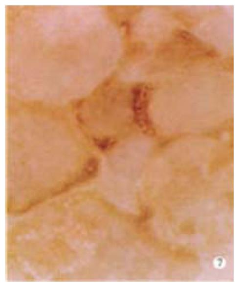
Figure 7 Immunohistochemical SP method, DAB staining and hematoxylin counter-staining cell nuclei.
Core antibody positive granules are seen as a brown lump with in the cytoplasm of LCL. × 400
- Citation: Cheng JL, Liu BL, Zhang Y, Tong WB, Yan Z, Feng BF. Hepatitis C virus in human B lymphocytes transformed by Epstein-Barr virus in vitro by in situ reverse transcriptase-polymerase chain reaction. World J Gastroenterol 2001; 7(3): 370-375
- URL: https://www.wjgnet.com/1007-9327/full/v7/i3/370.htm
- DOI: https://dx.doi.org/10.3748/wjg.v7.i3.370


