Copyright
©The Author(s) 1999.
World J Gastroenterol. Dec 15, 1999; 5(6): 492-505
Published online Dec 15, 1999. doi: 10.3748/wjg.v5.i6.492
Published online Dec 15, 1999. doi: 10.3748/wjg.v5.i6.492
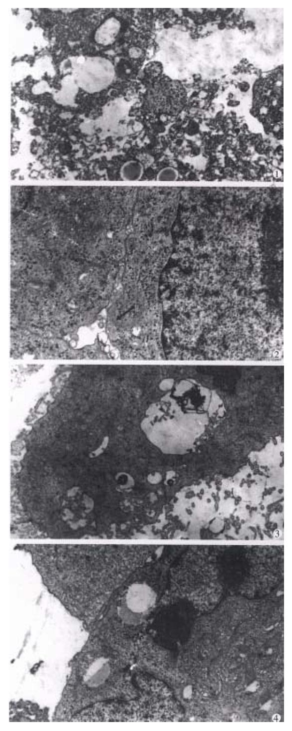
Figure 3 Results of intratumoral injection of 32P-GMS demonstrated by electron microscopy: ① Necrosed tumor cells ( × 5000 ).
② Formation of bile duct-like structure among tumor cells (solid arrow) ( × 8000). ③ Plenty of microvilli on the surface of tumor cell ( × 7000). ④ In control group, intratumoral injection of 32P-GMS showing heteromorphic tumor cell with cleavage of nucleus and scanty microvilli on surface ( × 7000).
- Citation: Liu L, Jiang Z, Teng GJ, Song JZ, Zhang DS, Guo QM, Fang W, He SC, Guo JH. Clinical and experimental study on regional administration of phosphorus 32 glass microspheres in treating hepatic carcinoma. World J Gastroenterol 1999; 5(6): 492-505
- URL: https://www.wjgnet.com/1007-9327/full/v5/i6/492.htm
- DOI: https://dx.doi.org/10.3748/wjg.v5.i6.492


