Copyright
©The Author(s) 2021.
World J Gastroenterol. Jun 14, 2021; 27(22): 3097-3108
Published online Jun 14, 2021. doi: 10.3748/wjg.v27.i22.3097
Published online Jun 14, 2021. doi: 10.3748/wjg.v27.i22.3097
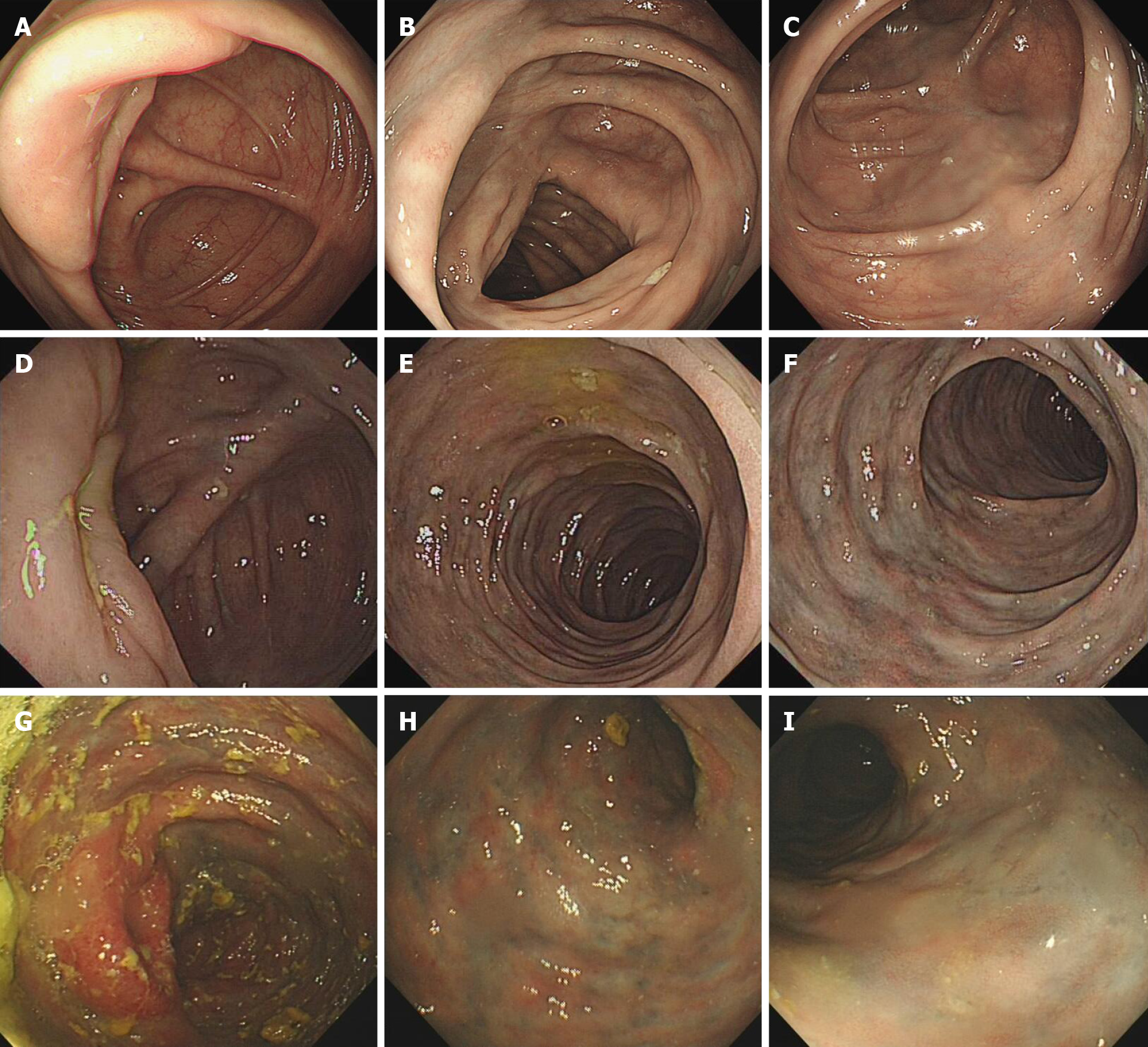
Figure 6 Representative endoscopic views.
A-C: Colonoscopy in case 3 revealed light blue discoloration in the transverse colon; D-F: Colonoscopy case 2 showed edematous congested mucosa with pigmentation, and dark blue discoloration extending to the transverse colon; G-I: Colonoscopy of case 5 revealed edematous dark purple colonic mucosa and sclerotic changes of the colonic walls extending from the cecum to the splenic flexure of colon.
- Citation: Wen Y, Chen YW, Meng AH, Zhao M, Fang SH, Ma YQ. Idiopathic mesenteric phlebosclerosis associated with long-term oral intake of geniposide. World J Gastroenterol 2021; 27(22): 3097-3108
- URL: https://www.wjgnet.com/1007-9327/full/v27/i22/3097.htm
- DOI: https://dx.doi.org/10.3748/wjg.v27.i22.3097


