Copyright
©The Author(s) 2020.
World J Gastroenterol. Nov 28, 2020; 26(44): 7076-7084
Published online Nov 28, 2020. doi: 10.3748/wjg.v26.i44.7076
Published online Nov 28, 2020. doi: 10.3748/wjg.v26.i44.7076
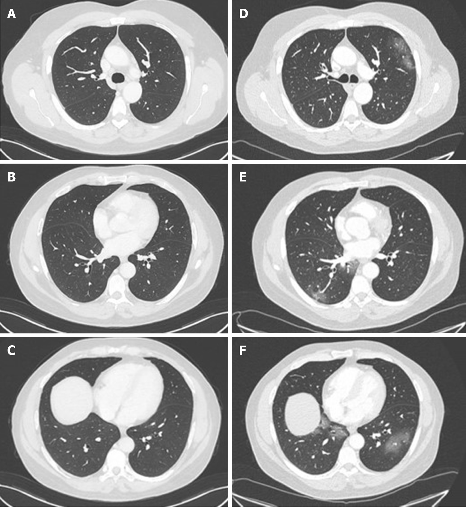
Figure 1 Chest computed tomography of a 58-year-old man who underwent liver transplantation in 2018.
A-C: Normal chest computed tomography (CT) of the patient in November 2019; D and E: Chest CT on admission in March 2020 showing ground-glass opacities with a peripheral distribution.
- Citation: Sessa A, Mazzola A, Lim C, Atif M, Pappatella J, Pourcher V, Scatton O, Conti F. COVID-19 in a liver transplant recipient: Could iatrogenic immunosuppression have prevented severe pneumonia? A case report. World J Gastroenterol 2020; 26(44): 7076-7084
- URL: https://www.wjgnet.com/1007-9327/full/v26/i44/7076.htm
- DOI: https://dx.doi.org/10.3748/wjg.v26.i44.7076


