Copyright
©The Author(s) 2019.
World J Gastroenterol. Dec 7, 2019; 25(45): 6693-6703
Published online Dec 7, 2019. doi: 10.3748/wjg.v25.i45.6693
Published online Dec 7, 2019. doi: 10.3748/wjg.v25.i45.6693
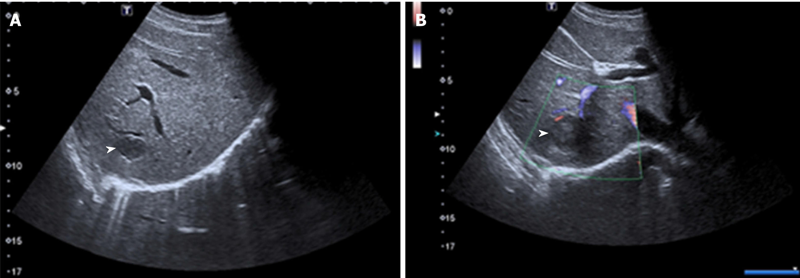
Figure 2 Unenhanced ultrasound images of Case 1.
A and B: Unenhanced ultrasound revealed two heterogeneous hypoechoic lesions without visualized internal blood flow. Inflammatory pseudotumor-like follicular dendritic cell tumors (black arrowheads) from the upper segment of the right posterior lobe were present in the liver of a 31-year-old Chinese woman.
- Citation: Li HL, Liu HP, Guo GWJ, Chen ZH, Zhou FQ, Liu P, Liu JB, Wan R, Mao ZQ. Imaging findings of inflammatory pseudotumor-like follicular dendritic cell tumors of the liver: Two case reports and literature review. World J Gastroenterol 2019; 25(45): 6693-6703
- URL: https://www.wjgnet.com/1007-9327/full/v25/i45/6693.htm
- DOI: https://dx.doi.org/10.3748/wjg.v25.i45.6693


