Copyright
©The Author(s) 2018.
World J Gastroenterol. Nov 7, 2018; 24(41): 4663-4678
Published online Nov 7, 2018. doi: 10.3748/wjg.v24.i41.4663
Published online Nov 7, 2018. doi: 10.3748/wjg.v24.i41.4663
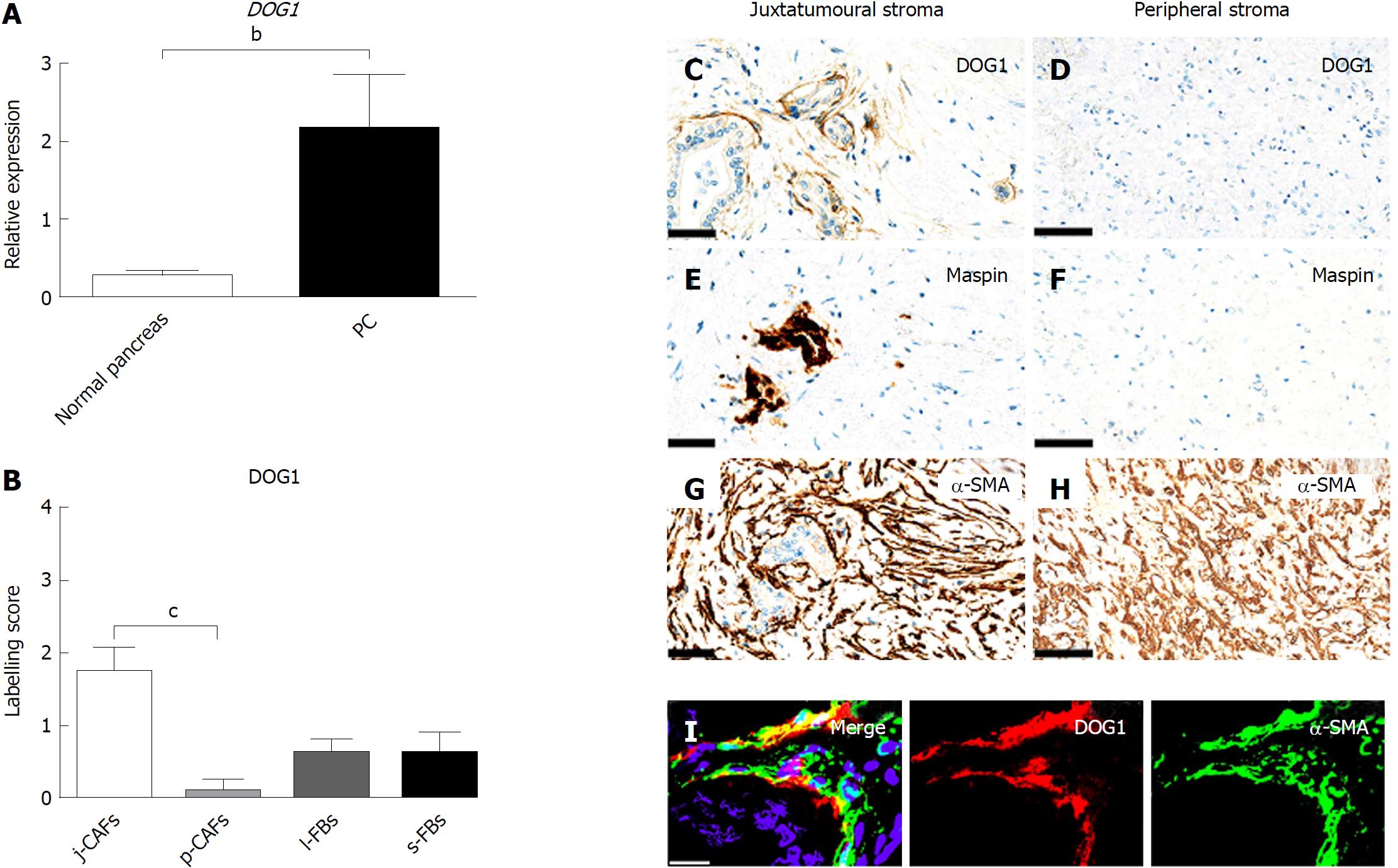
Figure 6 DOG1 gene and DOG1 protein expression in pancreatic cancer.
A: Expression of the DOG1 gene was significantly upregulated in pancreatic cancer compared to normal pancreatic specimens. bP < 0.01; B: Semi-quantitative mean labelling scores of DOG1 in juxtatumoural cancer-associated fibroblasts (j-CAFs) = 1.8, peripheral cancer-associated fibroblasts (p-CAFs) = 0.1, lobular fibroblasts = 0.6, and septal fibroblasts = 0.6. DOG1 is expressed at significantly higher levels in j-CAFs than in p-CAFs. cP < 0.001; C: Weak DOG1 expression in spindle-shaped cells in the juxtatumoural stroma. Some adenocarcinoma cells also expressed DOG1 (scale bar, 50 μm); D: No DOG1 expression was observed in the peripheral stroma (scale bar 100μm). Maspin-positive cancer cells are E: present in the juxtatumoural stroma (scale bar, 50 μm); but F: not in the peripheral stroma (scale bar, 100 μm). Strong α-smooth muscle actin (α-SMA) expression in G: j-CAFs (scale bar, 50 μm); and H: p-CAFs (scale bar, 100 μm); I: j-CAFs co-express DOG1 and α-SMA [double-immunofluorescence of α-SMA (green) and DOG1 (red); scale bar, 20 μm]. j-CAFs: Juxtatumoural cancer-associated fibroblasts; p-CAFs: Peripheral cancer-associated fibroblasts; l-FBs: Lobular fibroblasts; s-FBs: Septal fibroblasts; PC: Pancreatic cancer.
- Citation: Nielsen MFB, Mortensen MB, Detlefsen S. Typing of pancreatic cancer-associated fibroblasts identifies different subpopulations. World J Gastroenterol 2018; 24(41): 4663-4678
- URL: https://www.wjgnet.com/1007-9327/full/v24/i41/4663.htm
- DOI: https://dx.doi.org/10.3748/wjg.v24.i41.4663


