Copyright
©The Author(s) 2017.
World J Gastroenterol. Nov 14, 2017; 23(42): 7609-7617
Published online Nov 14, 2017. doi: 10.3748/wjg.v23.i42.7609
Published online Nov 14, 2017. doi: 10.3748/wjg.v23.i42.7609
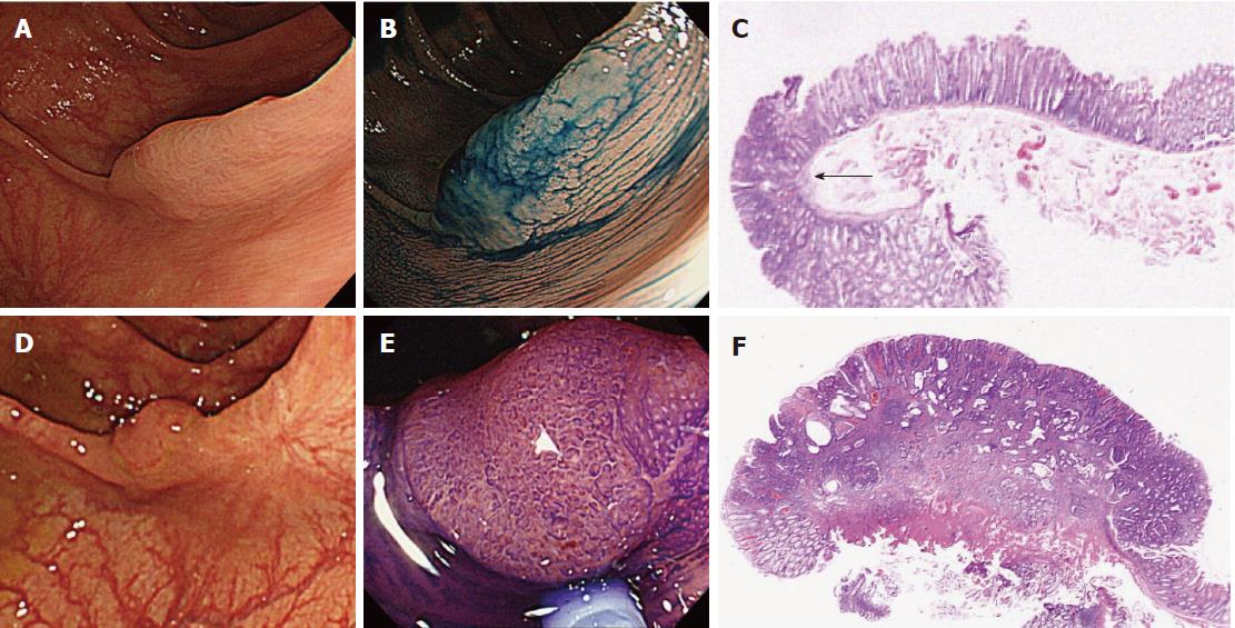
Figure 6 An early post-colonoscopy colorectal cancer case resulting from incomplete resection (No.
6 in Table 2). A 73-year-old woman underwent initial colonoscopy. A large flat lesion 20 mm in size showing type-II and open type-II pits, suggestive of SSA/P, was found by chromoendoscopy in the transverse colon and resected by piecemeal EMR with no macroscopically evident residual lesion (A and B); C: Histopathology of the resected specimen divided into 3 pieces revealed high-grade dysplasia (arrow) in SSA/P with intact vertical and horizontal margins of the dysplasia; D: Surveillance colonoscopy 11 months after initial colonoscopy detected a flat 10-mm lesion on the scar of the initial EMR in the transverse colon; E: Chromoendoscopy with crystal violet revealed unusual type-Vi pits suggesting submucosal invasive cancer; F: Histopathological examination of the EMR specimen revealed moderately differentiated adenocarcinoma invading the deep (2500 μm) submucosa with lymphovascular involvement. Finally, surgery was performed and histopathological examination revealed no residual cancer at the EMR site in the transverse colon and no lymph node metastasis. EMR: Endoscopic mucosal resection; SSA/P: Sessile serrated adenoma/polyp.
- Citation: Iwatate M, Kitagawa T, Katayama Y, Tokutomi N, Ban S, Hattori S, Hasuike N, Sano W, Sano Y, Tamano M. Post-colonoscopy colorectal cancer rate in the era of high-definition colonoscopy. World J Gastroenterol 2017; 23(42): 7609-7617
- URL: https://www.wjgnet.com/1007-9327/full/v23/i42/7609.htm
- DOI: https://dx.doi.org/10.3748/wjg.v23.i42.7609


