Copyright
©The Author(s) 2017.
World J Gastroenterol. Oct 14, 2017; 23(38): 7047-7053
Published online Oct 14, 2017. doi: 10.3748/wjg.v23.i38.7047
Published online Oct 14, 2017. doi: 10.3748/wjg.v23.i38.7047
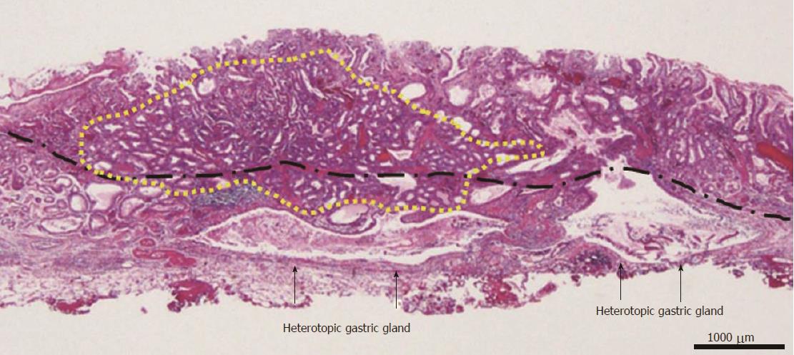
Figure 6 Histopathological findings of the endoscopic submucosal dissection-resected specimen.
Yellow line the gastric adenocarcinoma of fundic gland type (GA-FG), Black line muscularis mucosae. The GA-FG partially invaded the submucosa layer up to 450 μm, although it was located mainly in the deep layer of the lamina propria mucosae. Enlarged ducts of the heterotopic gastric glands were concomitantly observed just under the submucosal lesion of the GA-FG. In addition, the submucosal lesion had a poor stromal reaction.
- Citation: Manabe S, Mukaisho KI, Yasuoka T, Usui F, Matsuyama T, Hirata I, Boku Y, Takahashi S. Gastric adenocarcinoma of fundic gland type spreading to heterotopic gastric glands. World J Gastroenterol 2017; 23(38): 7047-7053
- URL: https://www.wjgnet.com/1007-9327/full/v23/i38/7047.htm
- DOI: https://dx.doi.org/10.3748/wjg.v23.i38.7047


