Copyright
©The Author(s) 2016.
World J Gastroenterol. May 28, 2016; 22(20): 4901-4907
Published online May 28, 2016. doi: 10.3748/wjg.v22.i20.4901
Published online May 28, 2016. doi: 10.3748/wjg.v22.i20.4901
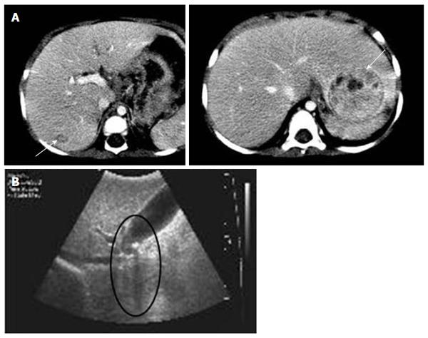
Figure 1 Radiologic hepatic evaluations in patient 1 and 2.
A: Abdominal computed tomography of patient 1 revealed two contrast-enhanced hepatic masses (arrows) at 21 mo of age; B: Gallstone and its posterior shadow (circle) were observed on liver ultrasonography in patient 2.
- Citation: Park JS, Ko JS, Seo JK, Moon JS, Park SS. Clinical and ABCB11 profiles in Korean infants with progressive familial intrahepatic cholestasis. World J Gastroenterol 2016; 22(20): 4901-4907
- URL: https://www.wjgnet.com/1007-9327/full/v22/i20/4901.htm
- DOI: https://dx.doi.org/10.3748/wjg.v22.i20.4901


