Copyright
©The Author(s) 2015.
World J Gastroenterol. Nov 28, 2015; 21(44): 12709-12712
Published online Nov 28, 2015. doi: 10.3748/wjg.v21.i44.12709
Published online Nov 28, 2015. doi: 10.3748/wjg.v21.i44.12709
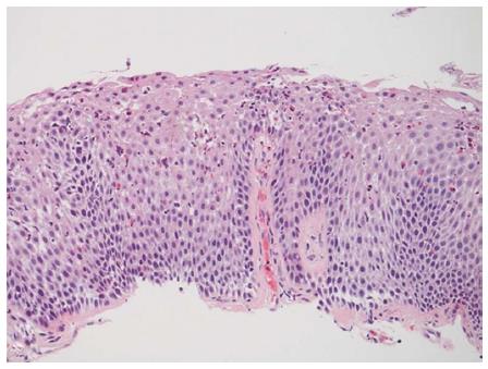
Figure 3 Esophageal (A), gastric (B), and duodenal (C) biopsies showing moderate to severe eosinophilic infiltration of the lamina propria and epithelium.
The duodenal biopsy also shows gastric metaplasia. Hematoxylin-eosin staining, magnification × 200.
- Citation: Riggle KM, Wahbeh G, Williams EM, Riehle KJ. Perforated duodenal ulcer: An unusual manifestation of allergic eosinophilic gastroenteritis. World J Gastroenterol 2015; 21(44): 12709-12712
- URL: https://www.wjgnet.com/1007-9327/full/v21/i44/12709.htm
- DOI: https://dx.doi.org/10.3748/wjg.v21.i44.12709


