Copyright
©The Author(s) 2015.
World J Gastroenterol. Nov 21, 2015; 21(43): 12249-12260
Published online Nov 21, 2015. doi: 10.3748/wjg.v21.i43.12249
Published online Nov 21, 2015. doi: 10.3748/wjg.v21.i43.12249
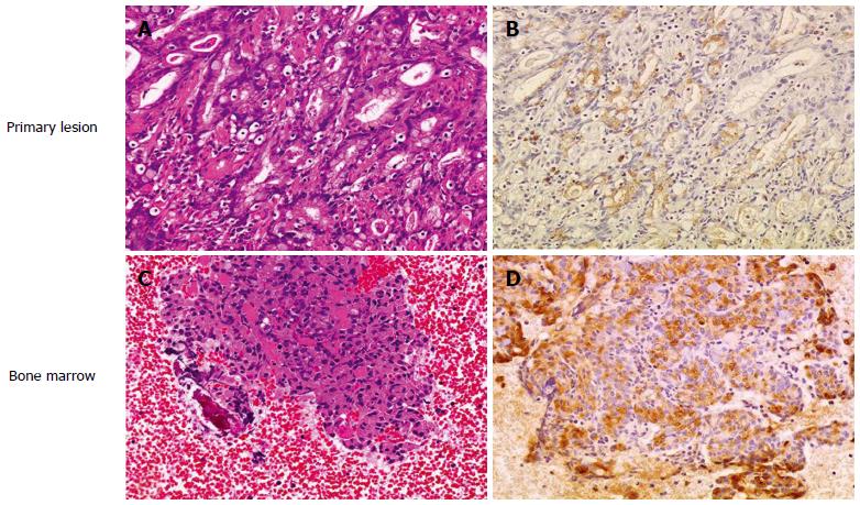
Figure 2 RANKL expression in disseminated carcinomatosis of the bone marrow associated with gastric cancer.
Representative immunohistochemistry for RANKL in gastric cancer demonstrating disseminated carcinomatosis of the bone marrow. A: Hematoxylin and eosin staining of gastric cancer shows moderately differentiated adenocarcinoma (magnification × 20); B: Immunohistochemistry for RANKL in a serial section of the same specimen in (A). RANKL shows positive staining predominantly in the cytoplasm and plasma membrane of moderately differentiated adenocarcinoma cells (magnification × 20); C: Hematoxylin and eosin staining of a bone marrow aspiration smear shows infiltration of atypical epithelial cells, indicating metastasis from known gastric cancer (magnification × 20); D: Immunohistochemistry for RANKL in a serial section of the same specimen in (C). RANKL shows positive staining predominantly in the cytoplasm and plasma membrane of metastatic gastric cancer cells as is seen in the primary lesion (B) (magnification × 20). Adapted from Kusumoto et al[9].
- Citation: Iguchi H. Recent aspects for disseminated carcinomatosis of the bone marrow associated with gastric cancer: What has been done for the past, and what will be needed in future? World J Gastroenterol 2015; 21(43): 12249-12260
- URL: https://www.wjgnet.com/1007-9327/full/v21/i43/12249.htm
- DOI: https://dx.doi.org/10.3748/wjg.v21.i43.12249


